
Page 155 - Read Online
P. 155
Ieni et al. Colonic metastasis by a uterine LCNEC
derived from pluripotent stem cells with the possibility such as chromogranin, synaptophysin and CD56. In
[5]
for both neuroendocrine and glandular endometrioid this case, the diagnosis of endometrial LCNEC was
differentiation. [11] based on neuroendocrine appearance, particularly the
neuroendocrine marker expression (synaptophysin
In the current literature, 15 cases of endometrial and partial CD56 reactivity). In differential diagnoses,
LCNEC have been described in patients with a endometrial NECs should be distinguished from other
mean age of 64 years, 8 of which cases are pure tumors characterized by nuclear high-grade features
and 7 are associated with another component. In with a predominantly solid growth pattern, such as
[8]
particular, the pure form LCNEC is characterized carcinosarcoma, undifferentiated endometrial sarcoma,
by solid sheets with organoid, trabecular or cord- solid pattern of serous carcinoma and undifferentiated
like patterns including peripheral palisading and endometrial carcinoma (UEC). However, the most
necrosis areas. The neoplastic cells have large problematic differential diagnosis is represented by
[8]
and abundant eosinophilic cytoplasm with vesicular UEC, in which a focal neuroendocrine differentiation
high-grade nuclei, prominent nucleoli and frequent (< 10%), with 1 or more neuroendocrine markers, has
mitotic figures. The confirmation of neuroendocrine been demonstrated in 41% in UEC series; therefore,
[9]
[12]
differentiation is based on neuroendocrine markers, the expression of neuroendocrine markers in more
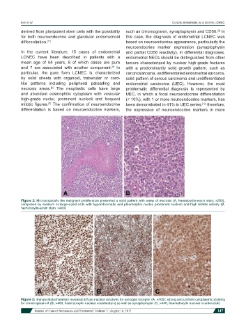
Figure 2: Microscopically the malignant proliferation presented a solid pattern with areas of necrosis (A, hematoxylin-eosin stain, ×200),
composed by medium or large-sized cells with hyperchromatic and pleomorphic nuclei, prominent nucleoli and high mitotic activity (B,
hematoxylin-eosin stain, ×400)
Figure 3: Immunohistochemistry revealed diffuse nuclear reactivity for estrogen receptor (A, ×400); strong and uniform cytoplasmic staining
for chromogranin-A (B, ×400, haematoxylin nuclear counterstain) as well as synaptophysin (C, ×400, haematoxylin nuclear counterstain)
Journal of Cancer Metastasis and Treatment ¦ Volume 3 ¦ August 16, 2017 147