
Page 149 - Read Online
P. 149
Agrawal et al. Challenging treatment of huge fibromatosis
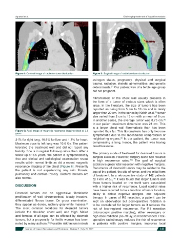
Figure 4: Coronal image of radiation dose distribution Figure 5: Sagittal image of radiation dose distribution
estrogen status, pregnancy, physical and surgical
trauma, radiation, skeletal abnormalities, and genetic
determinants. Our patient was of a fertile age group
[7]
but not pregnant.
Fibromatosis of the chest wall usually presents in
the form of a tumor of various sizes which is often
large. In the literature, the size of tumors has been
reported as being from 5 cm to 10 cm and is rarely
larger than 20 cm. In the series by Kabiri et al. tumor
[3]
size varied from 2 cm to 13 cm with a mean of 6 cm.
In another series, the average tumor was 8.75 cm.
[8]
In our patient maximum dimension was 21 cm. This
is a larger chest wall fibromatosis than has been
Figure 6: Axial image of magnetic resonance imaging chest at 3.5 reported thus far. This fibromatosis has only become
years
symptomatic due to the mechanical compression of
[9]
21% for right lung, 16.6% for liver and 1.8% for heart. neighboring organs. In our patient, the tumor was
Maximum dose to left lung was 10.6 Gy. The patient compressing a lung, hence, the patient was having
tolerated the treatment well and did not report any breathlessness.
toxicity. She is in regular follow-up since then. After a
follow-up of 3.5 years, the patient is symptomatically The primary mode of treatment for desmoid tumors is
free and clinical and radiological examination reveal surgical excision. However, surgery alone has resulted
in high recurrence rates. The goal of surgical
[10]
results within normal limits as did a recent magnetic excision is gross total resection with negative margins.
resonance imaging of the chest [Figure 6]. Presently Recurrence of desmoid tumors may be related to the
the patient is not experiencing any skin fibrosis, age of the patient, the site of tumor, and the initial form
pulmonary and cardiac toxicity. Bilateral breasts are of treatment. In a retrospective study of 142 patients
also normal. by Fiore et al., it was found that larger tumors and
[11]
those tumors located on the trunk were associated
DISCUSSION with a higher risk of recurrence. Local control rates
have been reported to be a function of tumor location,
Desmoid tumors are an aggressive fibroblastic ability to obtain negative margins, and adjuvant
proliferation of well circumscribed, locally invasive, therapy. In cases of R0 resection, a patient can be
differentiated fibrous tissue. On gross examination, kept on observation but post-operative radiation is
they appear as dense, rubbery gray-white masses. to be considered for larger tumors as it reduces the
[5]
The most common locations for desmoid tumors risk of loco-regional recurrence. For R1 resection
include the shoulder, chest wall, and thigh. Males (microscopic positive margins), either re-surgery or
and females of all ages can be affected by desmoid high dose radiation (66-70 Gy) is recommended. Post-
tumors, but a propensity for fertile women has been operative radiotherapy reduces the risk of recurrence
noted by many authors. Possible risk factors include in patients with positive margins, improves local
[6]
Journal of Cancer Metastasis and Treatment ¦ Volume 3 ¦ July 21, 2017 141