
Page 141 - Read Online
P. 141
to unusual sites with myriad presentations. Of the RCC rarely associated with prodigious metastasis. This has been
sub-types, clear cell RCC is notorious for its unpredictable attributed to its hypovascular nature, owing to the lack
metastatic pattern; on the other hand, papillary RCC is of Von Hippel-Lindau mutations that regulate vascular
endothelial growth factor, the primary proangiogenic
molecule in RCC. The relative rarity of papillary RCC
[1]
metastatic to the bladder was also demonstrated in a recent
series of 11 cases of metastatic RCC to the urinary
bladder that were detected over a span of 15 years, with
only 18% (2/11) originating from papillary RCC. [2]
The bladder is an unusual site for metastasis of RCC with
an incidence of 1.6% in autopsy series. Other metastatic
[3]
sites of RCC to the genitourinary tract include the ipsilateral
ureter, contralateral ureter, ureteric stump and prostatic
fossa. Bladder metastasis may be solitary or multiple,
the latter having a worse prognosis. Both synchronous and
metachronous bladder metastasis from RCC have been
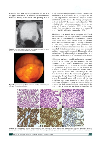
Figure 1: Contrast-enhanced computed tomography: Moderately enhancing described. Metachronous lesions occur more commonly
lesion in the left postero-inferior wall of the bladder and have been reported to occur up to 12 years after radical
nephrectomy. Synchronous lesions are more likely to be
[4]
associated with the presence of metastasis in other organs.
Although a variety of possible pathways for metastasis
of RCC to the bladder have been proposed, the exact
mechanism is not clear. Hematogenous spread may
[5]
occur through the general circulation or retrograde through
the periureteric or gonadal veins. In this scenario, the
metastasis is usually located within the bladder detrusor
layer. Lymphatic spread may occur through the renal
hilar lymphatics down the periureteral lymphatics and
subsequently through the pelvic lymphatics to the pelvic
organs. Transluminal spread with seeding of the distal
urothelium may occur, especially in cases where the
renal tumor infiltrates the pelvicalyceal system. We believe
Figure 2: Cystoscopic image: Broad-based non-papillary lesion arising from this to be the likely mechanism in our patient, considering
the region of the left ureteric orifice that the site of metastasis was in the region of the left
Figure 3: (a-c) Histopathology showing papillary adenocarcinoma with moderate nuclear pleomorphism and eosinophilic cytoplasm. Hematoxylin and Eosin
staining section (a, ×4; b, ×40); (d) Immunohistochemistry shows strong positivity for cytokeratin 7; (e)vimentin; (f) focal positivity for cluster of differentiation 10
Journal of Cancer Metastasis and Treatment ¦ Volume 2 ¦ March 25, 2016 ¦ 131