
Page 144 - Read Online
P. 144
imaging methods based only on morphology and size. a 360° arc, using a low-energy, general purpose collimator.
Functional nuclear medicine imaging has the unique Acquisition was obtained with a matrix size of 64 × 64
advantage of assessment of the metabolic state of lymph × 16, 1.85 zoom factor, and a 15% symmetric window at
nodes. Currently, the agents employed are Thalium-201 140 keV (no contour). Reconstruction method was filtered
( Tl) and the two technetium-99m labelled compounds back projection (filter butterworth, cut-off frequency 0.5,
201
Sestamibi (MIBI) and Tetrofosmin. [2] power 7.0). No attenuation correction was used. Finally,
99m Tc-MIBI and Tc99m-tetrofosmin ( 99m Tc-TF) are two whole body scan was acquired in all patients for possible
lipophilic cationic complexes, which were originally distant metastases evaluation.
employed in myocardial perfusion imaging, but later were
found to possess tumor-seeking properties in the evaluation Two nuclear medicine specialists visually evaluated the
of diverse human malignancies. The diagnostic value planar, tomographic and whole body images, which were
[12]
of 99m Tc-TF could hold promise as a head and neck cancer compared to the CT scans. Increased uptake in SPECT
tracer, although limited data exist in clinical research. [13,15] images (in a site of pathological finding on CT (primary
In the present study the diagnostic utility of 99m Tc-TF prior tumor or lymph node) was considered a positive finding. A
to surgery of head and neck neoplasms was assessed and region of interest (ROI) was drawn on the relative coronal
correlated with the 99m Tc-TF uptake of histological grade, image. Background (Bg) ROI was drawn over the apex of
and tumor and lymph node size. the right lung. T/Bg index for tumor and lymph nodes was
METHODS derived in all patients with positive findings. Patients with
no significant uptake on pathological sites were considered
Prior to surgery, 12 subjects (11 males and 1 female) negative.
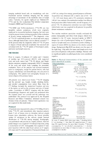
of median age 65.5-year-old (48-83) with suspected Table 1: Physical characteristics of the patients and
head and neck cancer had 99m Tc-TF planar and Single tumor anatomical location
photon emission computer tomography (SPECT) imaging Characteristics Patients
of the neck and whole body scanning for metastatic Gender 11 M/1 F
evaluation. None of the patients had received treatment Mean age (years) 65.75 ± 11.8 (48-83)
prior to scintigraphy, except for patient twelve, who had Height (cm) 166.75 ± 8.9 (155-184)
69.25 ± 9.5 (55-82)
Weight (kg)
surgery, radiotherapy and chemotherapy, one year prior to BMI (kg/m ) 25.00 ± 3.8 (16.2-32.4)
2
scintigraphy. This patient had scintigraphy because of a Smoking 11/12 (91.7%)
tumor relapse at the primary tumor.
* 0: 1/12 (8.3%)
All patients were interviewed before 99m Tc-TF imaging * 1: 0/12 (0.0%)
and the patients’ age, height, weight, smoking habits,
and alcohol consumption were noted. Data from the * 2: 11/12 (91.7%)
CT findings included the anatomical location and size Alcohol 8/12 (66.7%)
of the tumor, as well as the existence and size of lymph ** 0: 4/12 (33.3%)
nodes. Correlation of SPECT imaging with the tumor
histopathology diagnosis and grade was performed after ** 1: 2/12 (16.7%)
the original tumor was excised and the pathology was
established. The protocol was approved by the Hospital ** 2: 6/12 (50.0%)
Research and Ethics Committee. Informed consent was Anatomical location of
tumor
obtained from all patients. Clinical trial registration was Eepiglottis 1/12 (8.3%)
not required for this small study. Vocal cord 2/12 (16.7%)
Base of tongue 2/12 (16.7%)
Protocol Pharyngeal wall 1/12 (8.3%)
1/12 (8.3%)
Tonsil
All patients were injected with a dose of 740 MBq (20 mCi) Submandibular gland 1/12 (8.3%)
1/12 (8.3%)
bolus 99m Tc-TF; and immediately after injection patients Soft palate 2/12 (16.7%)
Pyriform fossa
drank 5 mL of lemon juice to stimulate salivary glands. Nasopharynx 1/12 (8.3%)
Lemon juice stimulation achieves the lowest possible Histopathological findings
uptake of radiotracer in the salivary glands (normal Squamous cell 10/12 (83.3%)
2/12 (16.7%)
Adenoid cystic cell
distribution of 99m Tc-TF) during imaging. Scintigraphy M: male; F: female; BMI: body mass index; * Smoking habits:
was performed with patients in a supine position. Anterior 0 = no smoking; 1 = less than 500 cigarettes/year; 2 = more
planar images were acquired 5-10 min post injection, using than 500 cigarettes/year; for more than 2 decades. **Alcohol
a zoom factor of 2.66. Tomography was acquired 15 min consumption: 0 = no alcohol consumption, 1 = alcohol
post-injection with the dual-head camera at 6 -angles consumption no more than 2-3 times in week; 2 = every day
o
(60 stops) and 30 s per projection (30 projection/head) over consumption of alcohol
134
Journal of Cancer Metastasis and Treatment ¦ Volume 2 ¦ April 15, 2016 ¦