
Page 138 - Read Online
P. 138
missing. Orthopantomograph revealed the presence of an procedure and was referred to the radiotherapy center
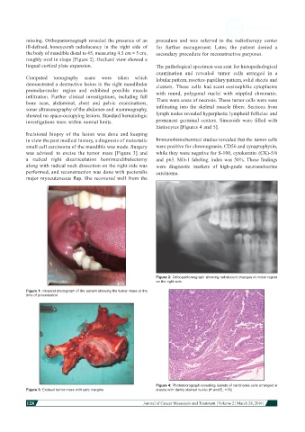
ill-defined, honeycomb radiolucency in the right side of for further management. Later, the patient denied a
the body of mandible distal to 45, measuring 4.5 cm × 5 cm, secondary procedure for reconstructive purposes.
roughly oval in shape [Figure 2]. Occlusal view showed a
lingual cortical plate expansion. The pathological specimen was sent for histopathological
examination and revealed tumor cells arranged in a
Computed tomography scans were taken which lobular pattern, rosettes-papillary pattern, solid sheets and
demonstrated a destructive lesion in the right mandibular clusters. These cells had scant eosinophilic cytoplasms
premolar-molar region and exhibited possible muscle with round, polygonal nuclei with stippled chromatin.
infiltration. Further clinical investigations, including full There were areas of necrosis. These tumor cells were seen
bone scan, abdominal, chest and pelvic examinations,
sonar ultrasonography of the abdomen and mammography, infiltrating into the skeletal muscle fibers. Sections from
showed no space-occupying lesions. Standard hematologic lymph nodes revealed hyperplastic lymphoid follicles and
investigations were within normal limits. prominent germinal centers. Sinusoids were filled with
histiocytes [Figures 4 and 5].
Incisional biopsy of the lesion was done and keeping
in view the past medical history, a diagnosis of metastatic Immunohistochemical studies revealed that the tumor cells
small cell carcinoma of the mandible was made. Surgery were positive for chromogranin, CD56 and synaptophysin,
was advised to excise the tumor mass [Figure 3] and while they were negative for S-100, cytokeratin (CK)-5/6
a radical right disarticulation hemimandibulectomy and p63. Mib-1 labeling index was 50%. These findings
along with radical neck dissection on the right side was were diagnostic markers of high-grade neuroendocrine
performed, and reconstruction was done with pectoralis carcinoma.
major myocutaneous flap. She recovered well from the
Figure 2: Orthopantomograph showing radiolucent changes in molar region
on the right side
Figure 1: Intraoral photograph of the patient showing the tumor mass at the
time of presentation
Figure 4: Photomicrograph revealing islands of carcinoma cells arranged in
Figure 3: Excised tumor mass with safe margins sheets with darkly stained nuclei (H and E, ×10)
128
Journal of Cancer Metastasis and Treatment ¦ Volume 2 ¦ March 28, 2016 ¦