
Page 109 - Read Online
P. 109
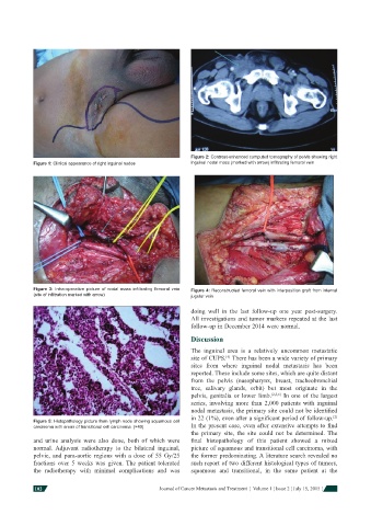
Figure 2: Contrast-enhanced computed tomography of pelvis showing right
            Figure 1: Clinical appearance of right inguinal nodes  inguinal nodal mass (marked with arrow) infi ltrating femoral vein


            Figure 3: Intra-operative picture of nodal mass infi ltrating femoral vein   Figure 4: Reconstructed femoral vein with interposition graft from internal
            (site of infi ltration marked with arrow)          jugular vein


                                                              doing well in the last follow-up one year post-surgery.
                                                              All investigations and tumor markers repeated at the last
                                                              follow-up in December 2014 were normal.

                                                              Discussion
                                                              The inguinal area is a relatively uncommon metastatic
                                                              site of CUPS.  There has been a wide variety of primary
                                                                          [4]
                                                              sites from where inguinal nodal metastasis has been
                                                              reported. These include some sites, which are quite distant
                                                              from the pelvis (nasopharynx, breast, tracheobronchial
                                                              tree, salivary glands, orbit) but most originate in the
                                                              pelvis, genitalia or lower limb. [2,5,6]  In one of the largest
                                                              series, involving more than 2,000 patients with inguinal
                                                              nodal metastasis, the primary site could not be identifi ed
                                                              in 22 (1%), even after a signifi cant period of follow-up.
                                                                                                            [2]
            Figure 5: Histopathology picture from lymph node showing squamous cell
            carcinoma with areas of transitional cell carcinoma. (×40)  In the present case, even after extensive attempts to fi nd
                                                              the primary site, the site could not be determined.  The
            and urine analysis were also done, both of which were   fi nal histopathology of this patient showed a mixed
            normal.  Adjuvant radiotherapy to the bilateral inguinal,   picture of squamous and transitional cell carcinoma, with
            pelvic, and para-aortic   regions with a dose of 55 Gy/25   the former predominating. A literature search revealed no
            fractions over 5 weeks was given.  The patient tolerated   such report of two different histological types of tumors,
            the radiotherapy with minimal complications and was   squamous and transitional, in the same patient at the


            102                                     Journal of Cancer Metastasis and Treatment  ¦  Volume 1 ¦ Issue 2 ¦ July 15, 2015 ¦