
Page 114 - Read Online
P. 114
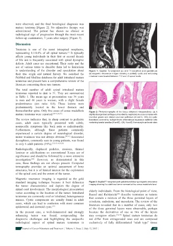
were observed, and the  fi nal histological diagnosis was
            mature teratoma [Figure 2]. No adjunctive therapy was
            administered.  The patient has shown no clinical or
            radiological sign of progression through the most recent
            follow-up examination, 3 years after surgery [Figure 3].
            Discussion
            Teratoma is one of the rarest intraspinal neoplasms,
            representing 0.1-0.6% of all spinal tumors.  It typically
                                               [1]
            affects young individuals in their  fi rst or second decade
            of life and is frequently associated with spinal dysraphic
            defects. Adult cases are exceptional.  Their rarity and the
            use of various terms to describe them led to limitations   a              b
            in understanding of the disorder and speculation about   Figure 1: Sagittal  T2-weighted (a) and  T1-weighted post-gadolinium
            their true origin and natural history.  We searched the   (b) magnetic resonance images showing a partially cystic and enhancing
            PubMed and Medline databases for adult intradural mature   intradural mass located between T12 and L3 spinal levels
            teratomas and present here a comprehensive review of the
            literature concerning these rare tumors.

            The total number of adult spinal intradural mature
            teratomas reported to date is 31.    They are summarized
            in  Table 1.  The mean age at presentation was 36 years
            in men and 44 years in women, with a slight female
            predominance (sex ratio: 0.8).  These lesions were
            predominantly located in the lower thoracic and    a                       b
            thoracolumbar spine. Only fi ve cases of conus medullaris   Figure 2: Photomicrographs of the tissue obtained intraoperatively. (a) A
            mature teratoma were reported. [14-16,26,27]      slightly disorganized cartilage surrounded by respiratory mucosa complete with
                                                              bronchial glands and ciliated columnar epithelium (H and E, ×20); (b) cystic
            The review indicates that, in sharp contrast to pediatric   formations covered by multiple levels of keratinous squamous epithelial cells
            cases, adult cases typically presented with subtle,   containing keratin lamellae (H and E, ×20). H and E: Hematoxylin and eosin stain
            nonspecifi c symptoms like back pain or radiculopathy.
            Furthermore,  although  these  patients  commonly
            experienced a certain degree of neurological disorder,
            motor weakness was not always obvious. [30,31]  Associated
            dysraphism, commonly seen in young patients, was found
            in only 6 adult patients (19%). [3,5,13,14,21,22]
            Radiologically, displaced pedicles, erosions, thinned
            laminae or calcifi cations on conventional X-rays are of
            signifi cance and should be followed by a more extensive
            investigation.  However, as demonstrated in this
                       [21]
            case, these  fi ndings are not always present. Computed
            tomography provides an optimal assessment of bony
            structures, but it is of limited interest for the exploration
            of the spinal cord and the extent of the tumor.
            Magnetic resonance imaging is regarded as the gold
            standard imaging technique because it best delineates   Figure 3: Sagittal T1-weighted post-gadolinium follow-up magnetic resonance
            the tumor characteristics and depicts the degree of   imaging showing the stabilized tumor remnant at the conus medullaris level
            spinal cord involvement. The morphological presentation   elderly individuals. From the histological point of view,
            varies according to the location of the tumor. Intradural   Russel and Rubinstein [32]  describe teratomas as tumors
            teratomas are commonly oval or lobulated, heterogeneous   that contain a mixture of the three germinal layers of
            masses. Cystic components are usually found in adult   ectoderm, endoderm, and mesoderm. The review of the
            cases, which can lead to confusion with more common   literature revealed that in a number of cases, only two
            epidermoid and dermoid cysts. [17]
                                                              of the three germinal layers were observable, perhaps
            In the present case, a well-demarcated and strongly   because the derivatives of one or two of the layers
            enhancing tumor was found, compounding the        may overgrow others. [10,19]  Spinal mature teratomas do
            diagnostic challenges and highlighting the unspecifi c   not differ from extragonadal ones and are composed
            radiological aspect of spinal mature teratomas in   exclusively of fully differentiated “adult type” tissue

                Journal of Cancer Metastasis and Treatment  ¦  Volume 1 ¦ Issue 2 ¦ July 15, 2015 ¦       107