
Page 104 - Read Online
P. 104
breast cancer MDA-MB-231 cells in vitro and in nude estrogen-independent. The WS extract caused increases
mice, respectively. WS roots have been used in ayurvedic in the percentage of MDA-MB-231 cells in the sub-G1
medicine for their anti-infl ammatory, analgesic, phase, indicating that WS causes apoptosis. Withaferin A,
anticancer, and anti-stress properties. [7,8] These diverse one of the active compounds of WS, causes G (2)/M cell
effects are attributed to the presence of active steroidal cycle arrest, associated with modulation of cyclin B1,
compounds that are called withanolides. Our current p34(cdc2), and PCNA levels, decreases the levels of
[15]
data showed that the WS extract inhibited proliferation STAT3 and its phosphorylation at Tyr(705) and Ser(727),
and metastasis of MDA-MB-231 cells in vitro and in and alters expression levels of p53-mediated apoptotic
nude mice. This inhibition was greater than that caused markers-Bcl2, Bax, caspase-3, and cleaved PARP. [18]
by withaferin A. The difference in inhibition may
[16]
be attributed to the fact that the whole extract contains Results of our current mouse experiments are consistent
active ingredients that have a synergistic effect against with in vitro data. The WS extract, administered orally,
breast cancer cells. [7,17] Since MDA-MB-231 cells are inhibited formation and growth of MDA-MB-231 cell
“triple-negative” form estrogen-independent tumors xenografts in nude mice, indicating that the active
in vivo, the anti-proliferative effect of WS is apparently ingredients of the WS extract are bioavailable after
oral administration. [19] Six mice of the untreated group
developed tumor metastasis to the lung, whereas none
MCF 10A
of the treated mice showed such tumor metastases.
100
This effect may be attributed to inhibition of CCL2
in xenografted tumors after treatment with WS root
% Cells Viability 60 extract. These results are consistent with a previous
80
concerning the inhibition of CCL2 in animals.
study
[20]
Inhibition of CCL2/CCR2 signaling by anti-CCL2
40
MCF 10A
antibodies blocks recruitment of infl ammatory
20
monocytes, inhibits metastasis, and prolongs the
survival of tumor-bearing mice. Depletion of tumor
0
0 12.5 µg/ml 25 µg/ml 50 µg/ml 100 µg/ml
cell-derived CCL2 also inhibits metastatic seeding.
Concentration of WS
Moreover, CCL2 mediates development of cancer stem
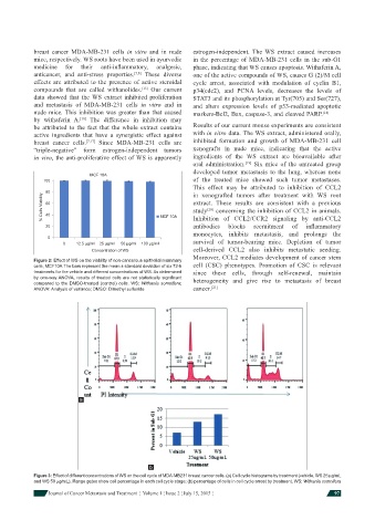
Figure 2: Effect of WS on the viability of non-cancerous epithelial mammary
cells, MCF10A.The bars represent the mean ± standard deviation of six 72-h cell (CSC) phenotypes. Promotion of CSC is relevant
treatments for the vehicle and different concentrations of WS. As determined since these cells, through self-renewal, maintain
by one-way ANOVA, results of treated cells are not statistically signifi cant heterogeneity and give rise to metastasis of breast
compared to the DMSO-treated (control) cells. WS: Withania somnifera;
ANOVA: Analysis of variance; DMSO: Dimethyl sulfoxide cancer. [21]
a
b
Figure 3: Effect of different concentrations of WS on the cell cycle of MDA-MB231 breast cancer cells. (a) Cell cycle histograms by treatment (vehicle, WS 25 μg/mL
and WS 50 μg/mL). Range gates show cell percentage in each cell cycle stage; (b) percentage of cells in cell cycle arrest by treatment. WS: Withania somnifera
Journal of Cancer Metastasis and Treatment ¦ Volume 1 ¦ Issue 2 ¦ July 15, 2015 ¦ 97