
Page 103 - Read Online
P. 103
(Agilent Technologies, Santa Clara, CA, USA), and Results
500 nM of each primer. β-Actin was used as the internal
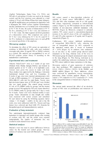
control, and the fi nal reactions were adjusted to a total WS extract caused a dose-dependent reduction of
volume of 20 μL with DNase RNase-free water (Qiagen). viability of breast cancer MDA-MB-231 cells by
All qPCR amplifi cation was performed in duplicates with 75% and 88% after treatment with 50 or 100 μg/mL
a Stratagene Mx 3005P system (Agilent Technologies), WS extract, respectively, compared to vehicle-treated
and the conditions were set to initial cycle of denaturation controls [Figure 1], but WS treatment did not affect the
at 95 °C for 10 min, 40 cycles of denaturation at 95 °C viability of non-cancerous epithelial mammary cells,
for 30 s, annealing at 55 °C for 1 min, and extension at MCF10A [Figure 2]. Moreover, compared to untreated
72 °C for 1 min. The fi nal segment involved generation controls, WS extract caused a concentration-dependent
of a dissociation curve. This comprised one cycle at increase in the sub-G1 phase of the cell population, by
95 °C for 1 min, followed by 55 °C for 30 s and 95 °C 6% and 10% after exposure to 25 μg/mL and 50 μg/mL,
for 30 s. Inclusion of a dissociation curve in each qPCR respectively [Figure 3].
run ensured specifi city of the amplicon. Furthermore, WS extract inhibited proliferation
Microarray analysis of xenografted MDA-MB-231 cells, reducing the
size of xenografted tumors by 60% compared to
To determine the effect of WS extract on expression of the untreated control after 8 weeks of treatment
cytokines in MDA-MB-231 cells, cells were incubated (P < 0.05) [Figure 4]. In addition, after euthanasia,
overnight with either 50 μg/mL WS or DMSO (vehicle) six of ten mice in the control group showed tumor
as a control. The analysis was accomplished by use of metastasis to the lung, whereas none of the mice in
HCA-II cytokine primer library II according to the WS-treated group developed metastasized tumor lesions
manufacturer’s instructions. in the lung [Figure 5]. This fi nding motivated us to
Experimental mice and treatments explore the underlying molecular mechanism by which
the WS extract inhibited tumor metastases to the lung.
nu
Athymic Nude-Foxn1 mice at 6 weeks of age were
obtained from Harlan Sprague-Dawley and housed in Microarray analysis of gene expression of cytokines
animal quarters at 22 °C with a 12 h light/dark cycle. was then performed. WS suppressed expression of
Animals were given free access to water and food. CCL2, CXCL1, CXCL2, CXCL3, IL1B, TGFB3, and
These studies were approved by the Tuskegee University BMP4 mRNA [Figure 6]. These inhibitory effects
Institutional Animal Care and Use Committee. At were confi rmed by quantitative reverse transcription-
8 weeks of age, mice were injected subcutaneously with polymerase chain reaction analysis [Figure 7]. WS
6
0.2 mL of PBS containing 1.5 × 10 human breast cancer caused a 75% reduction in CCL2 expression (P < 0.05)
MDA-MB-231 cells into the right fl anks. Twenty mice in the xenografted tumors of treated mice [Figure 8].
that developed tumor sizes of 50-200 mm were divided Discussion
3
into two equal groups. The control group received
0.2 mL of 5% DMSO orally by gavage, and the treated The current study assessed the effect of an alcoholic
group received 300 mg/kg/day WS root extract dissolved extract of WS roots on proliferation and metastasis of
in 5% DMSO orally by gavage daily for 5 days a week
for 8 weeks. Tumor sizes were checked weekly in each
120
group. Tumor dimensions in mm (length and width)
were measured with vernier calipers and calculated MDA-MB-231
for each tumor by using the following equation : tumor 100
volume = 1/2 (length × width ). At the end of the 8th
2
week, mice were euthanized with CO . Tumors and lung 80
2
tissues were collected and fi xed with 10% formalin for % Cell Viability MDA-MB-231
histopathological and immunochemistry analysis. 60
Evaluation of lung metastasis 40
Two pathologists histopathologically evaluated lung
metastases in untreated and treated groups after staining 20
of sections with HE, and the results were reported
independently. The number of metastatic foci was 0 0 12.5 µg/mL 25 µg/mL 50 µg/mL 100 µg/mL
counted in each stained tissue section. Concentration of WS
Statistical analyses Figure 1: Effect of WS on viability of breast cancer MDA-MB-231 cells. The
bars represent the mean ± standard deviation of six 24-h treatments for
Student’s t-test was used to assess differences between the vehicle and different concentrations of WS. The results are statistically
values for the treated and control groups. One-way signifi cant (P < 0.05) compared to the DMSO-treated (control) cells as
determined by one-way ANOVA with Dunnett’s test. WS: Withania somnifera;
analysis of variance was used with Dunnett’s test. ANOVA: Analysis of variance; DMSO: Dimethyl sulfoxide
96 Journal of Cancer Metastasis and Treatment ¦ Volume 1 ¦ Issue 2 ¦ July 15, 2015 ¦