
Page 13 - Read Online
P. 13
Page 8 of 18 Cheng et al. J Cancer Metastasis Treat 2021;7:17 https://dx.doi.org/10.20517/2394-4722.2021.27
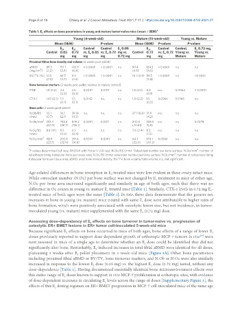
Table 1. E effects on bone parameters in young and mature tumor-naive mice (mean ± SEM) a
2
Young (4-week-old) Mature (15-week-old) Young vs. Mature
Mean (SEM) P-values Mean (SEM) P-values P-values
E , E , Control Control E 0.05 E , Control Control, E 0.72 mg,
2
2
2
2
2
Control 0.05 0.72 vs. E 0.05 vs. E 0.72 mg vs. Control 0.72 vs. E 0.72 Young vs. Young vs.
2
2
2
mg mg mg mg 0.72 mg mg mg Mature Mature
Proximal tibiae bone density and volume (6 weeks post-pellet)
aBMD 89.3 151.1 143.9 < 0.0001 < 0.0001 n.s. 99.4 134.3 < 0.0001 n.s. n.s.
2
(mg/cm ) (2.2) (2.9) (6.4) (4.8) (4.5)
BV/TV (%) 12.5 82.7 91.1 < 0.0001 < 0.0001 n.s. 10.1 (0.9) 59.5 < 0.0001 n.s. <0.0001
(2.0) (2.7) (2.4) (3.4)
Bone turnover markers (2 weeks post-pellet; relative to mature control)
P1NP 1.8 (0.2) 2.4 2.5 0.0401 0.0197 n.s. 1.0 (0.1) 0.8 n.s. 0.0064 < 0.0001
(0.2) (0.3) (0.1)
CTX-1 1.43 (0.1) 1.9 1.5 0.0142 n.s. n.s. 1.0 (0.2) 1.5 0.0066 0.0166 n.s.
(0.1) (0.1) (0.2)
Bone cells (2 weeks post-pellet)
N.Ob/BS 33.1 36.1 39.18 n.s. n.s. n.s. 27.1 (6.6) 31.4 n.s. n.s. n.s.
(mm) (0.7) (2.1) (4.3) (4.2)
2
N.Ob/mm 393.4 765.8 849.2 0.0051 0.0011 n.s. 240.0 528.3 n.s. n.s. 0.0278
(63.0) (86.3) (116.1) (70.61) (5.8)
N.Oc/BS 8.8 (1.1) 9.5 9.3 n.s. n.s. n.s. 7.6 (2.4) 8.3 n.s. n.s. n.s.
(mm) (0.6) (0.4) (1.5)
N.Oc/mm 2 112.9 202.0 217.4 0.0237 0.0097 n.s. 64.7 170.1 0.0263 n.s. n.s.
(22.7) (22.9) (14.6) (22.0) (47.2)
a 2
P-values determined by1-way ANOVA with Fisher’s LSD test. N.Ob/BS (mm): Osteoblast number per bone surface; N.Ob/mm : number of
2
osteoblasts lining trabecular bone per tissue area; N.Oc/BS (mm): osteoclast number per bone surface; N.Oc/mm : number of osteoclasts lining
trabecular bone per tissue area; aBMD: areal bone mineral density; BV/TV: bone volume/total volume; n.s.: not significant.
Age-related differences in bone resorption in E -treated mice were less evident in these ovary-intact mice.
2
While osteoclast number (N.Oc) per bone surface was not changed by E treatment in mice of either age,
2
N.Oc per bone area increased significantly and similarly in age of both ages, such that there was no
difference in Oc counts in young vs. mature E treated mice [Table 1]. Similarly, CTX-1 levels in 0.72 mg E -
2
2
treated mice of both ages were the same [Table 1]. In toto, these data demonstrate that the greater net
increases in bone in young (vs. mature) mice treated with same E dose were attributable to higher rates of
2
bone formation, which were positively associated with osteolytic lesion size, but not incidence, in tumor-
inoculated young (vs. mature) mice supplemented with the same E (0.72 mg) dose.
2
Assessing dose-dependency of E effects on bone turnover in tumor-naive vs. progression of
2
osteolytic ER+ BMET lesions in ER+ tumor cell-inoculated 5-week-old mice
Because significant E effects on bone occurred in mice of both ages, bone effects of a range of lower E
2
2
doses previously reported to support dose-dependent growth of orthotopic MCF-7 tumors in vivo were
[17]
next assessed in mice of a single age to determine whether an E dose could be identified that did not
2
significantly alter bone. Remarkably, E -induced increases in total tibial aBMD were identical for all doses,
2
plateauing 3 weeks after E -pellet placement in 5-week-old mice [Figure 3A]. Other bone parameters
2
including proximal tibial aBMD or BV/TV, bone turnover markers, and N.Ob or N.Oc were also similarly
increased in response to the lowest E dose (0.05 mg) vs. the highest E dose (0.72 mg) tested, without any
2
2
dose-dependence [Table 1]. Having documented essentially identical bone microenvironment effects over
this entire range of E doses known to support in vivo MCF-7 proliferation at orthotopic sites, with evidence
2
of dose-dependent increases in circulating E levels across the range of doses [Supplementary Figure 1], the
2
effects of this E dosing regimen on ER+ BMET progression in MCF-7 cell-inoculated mice of the same age
2