
Page 12 - Read Online
P. 12
Cheng et al. J Cancer Metastasis Treat 2021;7:17 https://dx.doi.org/10.20517/2394-4722.2021.27 Page 7 of 18
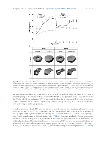
Figure 2. Effects of 0.72 mg E on bone mineral density and structure in tumor-naive young vs. skeletally mature mice. (A) Tibial areal
2
bone mineral density (BMD) in young (4-week-old) vs. skeletally mature (15-week-old) mice supplemented with 0.72 mg E (vs.
2
control), as measured by DXA (n = 6-8/group). Arrows indicate time of E pellet placement. *P ≤ 0.01, **P ≤ 0.0001 vs. age-matched
2
control, by 2-way ANOVA with Sidak post-test. (B) Representative microCT images of tibial cortical (top) and trabecular (bottom)
bone of young vs. skeletally mature E (0.72 mg)-supplemented mice (vs. age-matched controls), 6 weeks after E pellet placement.
2 2
confirmed dramatic, but differential, effects of E on both cortical and trabecular bone in the tibiae of
2
skeletally young vs. mature mice after 6 weeks of treatment. In the proximal tibial, a frequent osteolytic
BMET site, aBMD and trabecular BV/TV increased significantly in response to E in mice of both ages
2
[Table 1]; however, the increase was significantly greater in young mice [e.g., BV/TV of 91% vs. 60% (P <
0.0001) in young vs. mature, respectively].
In skeletally mature mice, P1NP, a serum marker of bone formation, was significantly lower (vs. young
mice) and unchanged by 0.72 mg E -supplementation, contrasting with a 0.72 mg E -induced increase in
2
2
already significantly higher P1NP levels in young mice, such that P1NP levels were 3.1-fold higher (P <
0.0001) in E -treated young vs. skeletally mature mice [Table 1]. Osteoblast number (N.Ob) per bone surface
2
tended to increase in response to E treatment in mice of both ages; however, these trends were not
2
statistically significant. Given the large increase in bone surface, N.Ob per area was also calculated and was
only significantly increased in response to E in young mice, resulting in Ob counts that were 1.6-fold higher
2
in young (vs. mature) E -treated mice (P < 0.05), consistent with higher circulating P1NP levels [Table 1].
2