
Page 11 - Read Online
P. 11
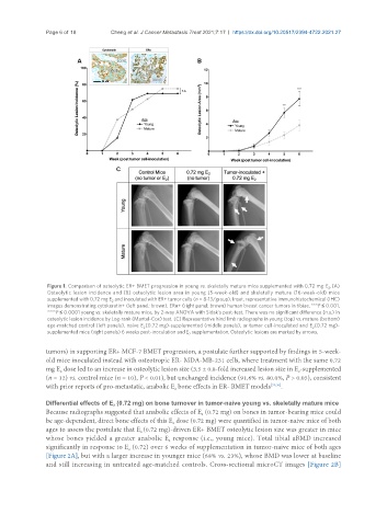
Page 6 of 18        Cheng et al. J Cancer Metastasis Treat 2021;7:17  https://dx.doi.org/10.20517/2394-4722.2021.27


                Figure 1. Comparison of osteolytic ER+ BMET progression in young vs. skeletally mature mice supplemented with 0.72 mg  E . (A)
                                                                                                      2
                Osteolytic lesion incidence and (B) osteolytic lesion area in young (5-week-old) and skeletally mature (16-week-old) mice
                supplemented with 0.72 mg E  and inoculated with ER+ tumor cells (n = 8-13/group). Inset, representative immunohistochemical (IHC)
                                   2
                images demonstrating cytokeratin+ (left panel; brown), ERα+ (right panel; brown) human breast cancer tumors in tibiae. ***P ≤ 0.001,
                ****P ≤ 0.0001 young vs. skeletally mature mice, by 2-way ANOVA with Sidak’s post-test. There was no significant difference (n.s.) in
                osteolytic lesion incidence by Log-rank (Mantel-Cox) test. (C) Representative hind limb radiographs in young (top) vs. mature (bottom)
                age-matched control (left panels), naive E (0.72 mg)-supplemented (middle panels), or tumor cell-inoculated and  E (0.72 mg)-
                                             2                                                   2
                supplemented mice (right panels) 6 weeks post-inoculation and E  supplementation. Osteolytic lesions are marked by arrows.
                                                           2
               tumors) in supporting ER+ MCF-7 BMET progression, a postulate further supported by findings in 5-week-
               old mice inoculated instead with osteotropic ER- MDA-MB-231 cells, where treatment with the same 0.72
               mg E  dose led to an increase in osteolytic lesion size (3.5 ± 0.8-fold increased lesion size in E -supplemented
                                                                                             2
                    2
               (n = 12) vs. control mice (n = 10), P < 0.01), but unchanged incidence (91.6% vs. 80.0%, P > 0.05), consistent
               with prior reports of pro-metastatic, anabolic E  bone effects in ER- BMET models [35,36] .
                                                       2
               Differential effects of E  (0.72 mg) on bone turnover in tumor-naive young vs. skeletally mature mice
                                    2
               Because radiographs suggested that anabolic effects of E  (0.72 mg) on bones in tumor-bearing mice could
                                                               2
               be age-dependent, direct bone effects of this E  dose (0.72 mg) were quantified in tumor-naive mice of both
                                                      2
               ages to assess the postulate that E (0.72 mg)-driven ER+ BMET osteolytic lesion size was greater in mice
                                            2
               whose bones yielded a greater anabolic E  response (i.e., young mice). Total tibial aBMD increased
                                                     2
               significantly in response to E  (0.72) over 6 weeks of supplementation in tumor-naive mice of both ages
                                         2
               [Figure 2A], but with a larger increase in younger mice (68% vs. 23%), whose BMD was lower at baseline
               and still increasing in untreated age-matched controls. Cross-sectional microCT images [Figure 2B]