
Page 381 - Read Online
P. 381
Page 10 of 16 Verkoeijen et al. J Cancer Metastasis Treat 2019;5:51 I http://dx.doi.org/10.20517/2394-4722.2019.06
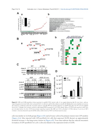
Figure 5. EGFR and EGFR-signaling is downregulated in paxillinS178A mutant cells. A: bar graph displaying the 10 most down- and up-
regulating genes in mutant cells when compared with wt cells (a); and Enrichr analysis using the GO Biological process function (processes
are sorted by combined score) (b); B: Enrichr analysis using the PPI Hub proteins function (a), EGFR being the first PPI with the highest
score (b); C: Western blot analysis confirms a decreased EGFR expression in all S178A clones. Ratios of EGFR levels relative to wt1 clone
are indicated below the blot. EGFR downstream signaling towards Akt, ERK and JNK is affected in S178A clones; D: activity of AP-1
member c-Jun was reduced in paxillinS178A cells as determined by TATA-luciferase reporter assay
cells was similar in the both groups [Figure 6Cb] and all tumor cells in the primary tumors were GFP-positive
[Figure 6Ca]. Mice injected with GFP-paxillinS178A cells that expressed EGFR showed an approximately
threefold increase in the lung tumor burden in [Figure 6D]. These data indicate that the reduced metastasis
formation of GFP-paxillinS178A cells is directly related to the expression levels of EGFR.