
Page 18 - Read Online
P. 18
Page 10 of 16 de Santis et al. Hepatoma Res 2019;5:1 I http://dx.doi.org/10.20517/2394-5079.2018.65
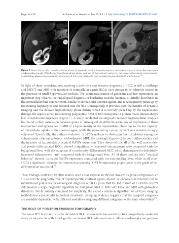
Figure 7. From left to right: hepatic arterial phase of gadoxetic acid-enhanced magnetic resonance imaging shows homogeneous
marked enhancement of the tumor; transitional phase shows washout of the contrast medium in the tumor with capsular enhancement;
hepatobiliary phase shows marked hypointensity of the tumor relative to the liver parenchyma. Modified from Park et al. [31]
In light of these considerations, reaching a definitive non invasive diagnosis of HCC is still a challenge
and MDCT and MRI with injection of extracellular agents (ECA) have proven to be relatively useless in
the presence of small hypovascular nodules. The commercialization of gadoxetic acid has represented an
important step towards the radiological diagnosis of borderline nodules because, it initially distributes in
the extracellular fluid compartment, similar to extracellular contrast agents, and is subsequently taken up by
functioning hepatocytes and excreted into the bile. Consequently, it provides both the benefits of dynamic
imaging and the delayed hepatobiliary phase during which it is actively picked up by the hepatocytes
through the organic anion transporting polypeptide (OATP) B1/8 transporter, a protein that is almost always
lost in hepatocarcinogenesis [Figure 7]. A study conducted on surgically resected hepatocellular nodules
has found a clear correlation between grade of histological de-differentiation, loss of expression of these
transporters and appearance in MRI of a hypointensity in the hepatobiliary phase due to the lost capacity
of intracellular uptake of the contrast agent, while the surrounding normal parenchyma remains strongly
enhanced. Specifically, the authors evaluated 72 HCCs nodules to determine the correlation among the
enhancement ratio on gadoxetic acid-enhanced MRI, the histological grade of tumour differentiation and
the intensity of immunohistochemical OATP8 expression. They observed that all of the well, moderately
and poorly differentiated HCCs showed a significantly decreased enhancement ratio compared with the
background liver, with the exception of 6 moderately differentiated HCC which demonstrated a definitively
increased enhancement ratio compared with the background liver. All of these nodules with “atypical
behavior” showed increased OATP8 expression compared with the surrounding liver, while in all other
HCCs a significant reduction in immunohistochemical OATP8 expression proportional to the grade of de-
[32]
differentiation was found .
These findings, confirmed by other studies, open a new scenario for the non-invasive diagnosis of hypovascular
HCCs but the diagnostic role of hepatospecific contrast agents should be endorsed and formalized in
international guidelines for radiological diagnosis of HCC, given that the last version of LI-RADS (v2017)
still provides a single diagnostic algorithm for multiphase MDCT, MRI with ECA, and MRI with gadoxetate
disodium. While initially combined for simplicity, the use of a common algorithm for all three imaging
methods has a potentially important drawback: emerging evidence suggests that the assigned categories
[33]
are modality-dependent, with different modalities assigning different categories to the same observation .
THE ROLE OF POSITRON EMISSION TOMOGRAPHY
The use of PET is still restricted in the field of HCC because of its low sensitivity. In a prospectively conducted
study on 99 patients with histologically confirmed HCC who underwent 18F-fluoro-deoxyglucose positron