
Page 21 - Read Online
P. 21
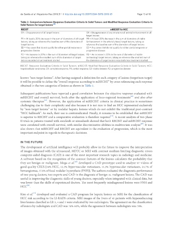
de Santis et al. Hepatoma Res 2019;5:1  I  http://dx.doi.org/10.20517/2394-5079.2018.65                                       Page 13 of 16


               Table 2. Comparison between Response Evaluation Criteria In Solid Tumors and Modified Response Evaluation Criteria In
               Solid Tumors for target lesions [41]
               RECIST                                                        mRECIST for HCC
               CR = Disappearance of all target lesions     CR = Disappearance of any intratumoral  arterial enhancement in all
                                                            target lesions
               PR = At least a 30% decrease in the sum of diameters of all target  PR = At least 30% decrease in the sum of diameters of viable
               lesions, taking as reference the baseline sum of the diameters of   (enhancement in the arterial phase) target lesions, taking as
               target lesions                               reference the baseline sum of the diameters of target lesions
               SD = Any cases that do not qualify for either partial response or   SD = Any cases that do not qualify for either partial response or
               progressive disease                          progressive disease
               PD = An increase in 20% in the sum of diameters of target lesions,  PD = An increase in 20% in the sum of diameters of viable
               taking as reference the smallest sum of the diameters of target   (enhancing) target lesions, taking as reference the smallest sum of
               lesions recorded since treatment started     the diameters of target lesions recorded since treatment started

               RECIST: Response Evaluation Criteria In Solid Tumors; mRECIST: Modified Response Evaluation Criteria In Solid Tumors; HCC:
               hepatocellular carcinoma; CR: complete response; PR: partial response; SD: stable disease; PD: progressive disease

               known “non target lesions”. After having assigned a definition for each category of lesions (target/non target)
               it will be possible to define the “overall response according to mRECIST” by cross-referencing each response
               obtained in the two categories of lesions as shown in Table 3.

               Subsequent publications have reported a good correlation between the objective response evaluated with
                                                                                           [42]
               mRECIST and overall survival, both after the application of loco-regional treatments  and also after
                               [43]
               systemic therapies . However, the application of mRECIST criteria in clinical practice is sometimes
               challenging due to their complexity and also because it is not rare to find an HCC represented exclusively
               by “non target lesions” or by nodular hepatic lesions which do not exhibit the traditional post-contrast
               “HCC hallmark”. As such, their use is contraindicated. Finally, it remains to be established that mRECIST
                                                                             [44]
               is superior to RECIST and a comparative evaluation is therefore required . A recent analysis of two phase
               II trials in patients treated with sorafenib or nintedanib showed that both RECIST and mRECIST response
                                                                                                  [45]
               were correlated with overall survival, with similar discriminative abilities in multivariate analysis . It was
               also shown that mRECIST and RECIST are equivalent in the evaluation of progression, which is the most
               important endpoint in regards to therapeutic decisions.


               IN THE FUTURE
               The development of artificial intelligence will probably allow in the future to improve the interpretation
               of images obtained with the ultrasound, MDTC or MRI with contrast medium limiting diagnostic errors
               computer-aided diagnosis (CAD) is one of the most important research topics in radiology and medicine.
               A software based on the recognition of the contrast features of the lesions calculates the probability that
                                                    [46]
               they are benign or malignant. Moga et al.  developed a CAD prototype used to analyze 97 videos of
               good quality CEUS [34% HCC, 12.3% hypervascular metastases, 11.3% hypovascular metastases, 24.7% of
               hemangiomas, 17.5% of focal nodular hyperplasia (FNH)]. The authors evaluated the diagnostic performance
               of two young doctors, two experts and CAD in the diagnosis of benign vs. malignant lesions. The CAD was
               useful in improving the diagnostic skills of young doctors, especially when integrated with clinical data, but
               was lower than the skills of experienced doctors. The most frequently misdiagnosed lesions were FNH and
                   [46]
               HCC .
                        [47]
               Kim et al.  developed and evaluated a CAD program for hepatic lesions on MRI for the classification of
               HCC risk according to the LI-RADS criteria. MRI images of the livers of 41 patients with hyperenhancing
               liver lesions classified as LR 3, 4 and 5 were evaluated by two radiologists. The agreement on the classification
               of lesions by radiologists and CAD was 76%-83%, while the agreement between radiologists was 78%.