
Page 26 - Read Online
P. 26
Page 2 of 9 Sukowati et al. Hepatoma Res 2019;5:2 I http://dx.doi.org/10.20517/2394-5079.2018.106
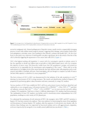
Figure 1. The oncogenicity of viral hepatitis in the development of hepatocellular carcinoma. HBV: hepatitis B virus; HCV: hepatitis C virus;
TGF: transforming growth factor; HCC: hepatocellular carcinoma; ER: endoplasmic reticulum
normal to malignant cells. Natural pathogenesis of hepatitis viruses usually involve a sequentially damaging
process. It starts with cellular immunological response, triggering DNA damage, mitochondrial dysfunction,
and endoplasmic reticulum stress, thus resulting in liver fibrosis, cirrhosis and finally HCC. On the other
hand, in the initial step, infection of viral hepatitis can play a significant role in the switch of the fate of the
cells by directly triggering the appearance of the cancer stem cells (CSC) [Figure 1].
CSC is the highest-ranking cell population in cancer with the tumorigenic capacity to initiate cancer. It
has the capability to divide and differentiate to partially or fully-differentiated cancer cells that comprise
the majority of cancer mass. This hierarchy model shows that CSC population is unique, with protective
[4]
mechanism to be responsible for the maintenance and propagation of the tumor . These cells act as the
main players in the highest level of the cancer hierarchy and may still have stem cells properties such as self-
renewal and ability to multiple cell types. Non-tumorigenic cells are thought to compose the bulk of tumors
[5,6]
but have little capacity to contribute to cancer progression .
[7,8]
The first evidence of CSC in HCC was demonstrated by the isolation of the side population in vitro
showing the involvement of CSC in drug resistance. The search and identification method of hepatic CSC
progressed by performing sphere colony formation and more commonly, by using CSC markers.
Various markers of CSC from established HCC cell lines and primary tumors had been identified and
validated by in vivo xenograft assay. Cell protein markers CD133 (PROM1) [9-11] , CD90 (THY-1) [12,13] , epithelial
[16]
cell adhesion molecule (EpCAM) [14,15] , CD24 , CD13 (ANPEP) [7,17] are the most common method to define
a hepatic CSC population. Until now, at least 12 different phenotypical CSC markers had been proposed.
The combination of these CSC markers was further used to characterize several subpopulations in a CSC
population, resulting in a wide variety of CSC phenotypes.
To understand the mechanism of early initiation of HCC, the oncogenic role of HBV and HCV proteins in
hepatic CSC has been started to be explored. They were analyzed by determining the extent of up-regulation
and the presence of various hepatic CSC markers, after the exposure of viral proteins into hepatic cells. In
addition, these findings were also supported by functional analysis such as cell aggressiveness, migration,
and more importantly, by xenograft in vivo model, several published data was shown in Table 1.