
Page 27 - Read Online
P. 27
Sukowati et al. Hepatoma Res 2019;5:2 I http://dx.doi.org/10.20517/2394-5079.2018.106 Page 3 of 9
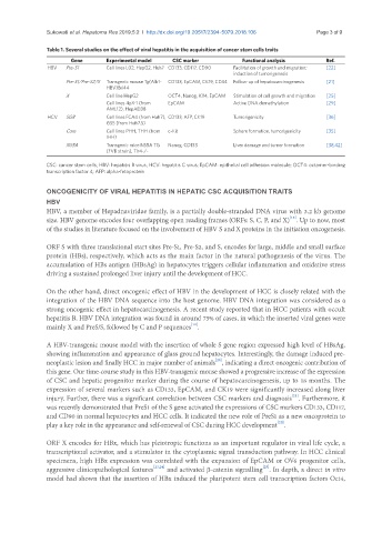
Table 1. Several studies on the effect of viral hepatitis in the acquisition of cancer stem cells traits
Gene Experimental model CSC marker Functional analysis Ref.
HBV Pre-S1 Cell lines L02, HepG2, Huh7 CD133, CD117, CD90 Facilitation of growth and migration; [22]
induction of tumorigenesis
Pre-S1/Pre-S2/S Transgenic mouse Tg(Alb1- CD133, EpCAM, CK19, CD34 Follow-up of hepatocarcinogenesis [21]
HBV)Bri44
X Cell line HepG2 OCT4, Nanog, Klf4, EpCAM Stimulation of cell growth and migration [25]
Cell lines 4pX-1 (from EpCAM Active DNA demethylation [29]
AML12), HepAD38
HCV SGR Cell lines FCA4 (from Huh7), CD133, AFP, CK19 Tumorigenicity [36]
GS5 (from Huh7.5)
Core Cell lines PHH, THH (from c-Kit Sphere formation, tumorigenicity [35]
IHH)
NS5A Transgenic mice NS5A TG Nanog, CD133 Liver damage and tumor formation [38,42]
(FVB strain), Tlr4-/-
CSC: cancer stem cells; HBV: hepatitis B virus; HCV: hepatitis C virus; EpCAM: epithelial cell adhesion molecule; OCT4: octamer-binding
transcription factor 4; AFP: alpha-fetoprotein
ONCOGENICITY OF VIRAL HEPATITIS IN HEPATIC CSC ACQUISITION TRAITS
HBV
HBV, a member of Hepadnaviridae family, is a partially double-stranded DNA virus with 3.2 kb genome
[18]
size. HBV genome encodes four overlapping open reading frames (ORFs: S, C, P, and X) . Up to now, most
of the studies in literature focused on the involvement of HBV S and X proteins in the initiation oncogenesis.
ORF S with three translational start sites Pre-S1, Pre-S2, and S, encodes for large, middle and small surface
protein (HBs), respectively, which acts as the main factor in the natural pathogenesis of the virus. The
accumulation of HBs antigen (HBsAg) in hepatocytes triggers cellular inflammation and oxidative stress
driving a sustained prolonged liver injury until the development of HCC.
On the other hand, direct oncogenic effect of HBV in the development of HCC is closely related with the
integration of the HBV DNA sequence into the host genome. HBV DNA integration was considered as a
strong oncogenic effect in hepatocarcinogenesis. A recent study reported that in HCC patients with occult
hepatitis B, HBV DNA integration was found in around 75% of cases, in which the inserted viral genes were
[19]
mainly X and PreS/S, followed by C and P sequences .
A HBV-transgenic mouse model with the insertion of whole S gene region expressed high level of HBsAg,
showing inflammation and appearance of glass ground hepatocytes. Interestingly, the damage induced pre-
[20]
neoplastic lesion and finally HCC in major number of animals , indicating a direct oncogenic contribution of
this gene. Our time-course study in this HBV-transgenic mouse showed a progressive increase of the expression
of CSC and hepatic progenitor marker during the course of hepatocarcinogenesis, up to 18 months. The
expression of several markers such as CD133, EpCAM, and CK19 were significantly increased along liver
[21]
injury. Further, there was a significant correlation between CSC markers and diagnosis . Furthermore, it
was recently demonstrated that PreS1 of the S gene activated the expressions of CSC markers CD133, CD117,
and CD90 in normal hepatocytes and HCC cells. It indicated the new role of PreS1 as a new oncoprotein to
[22]
play a key role in the appearance and self-renewal of CSC during HCC development .
ORF X encodes for HBx, which has pleiotropic functions as an important regulator in viral life cycle, a
transcriptional activator, and a stimulator in the cytoplasmic signal transduction pathway. In HCC clinical
specimens, high HBx expression was correlated with the expansion of EpCAM or OV6 progenitor cells,
[25]
aggressive clinicopathological features [23,24] and activated β-catenin signalling . In depth, a direct in vitro
model had shown that the insertion of HBx induced the pluripotent stem cell transcription factors Oct4,