
Page 15 - Read Online
P. 15
de Santis et al. Hepatoma Res 2019;5:1 I http://dx.doi.org/10.20517/2394-5079.2018.65 Page 7 of 16
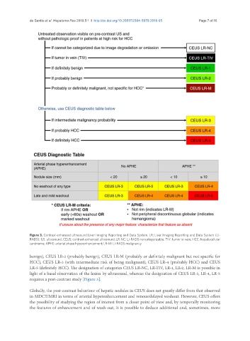
Figure 3. Contrast-enhanced ultrasound Liver Imaging Reporting and Data System. LR: Liver Imaging Reporting and Data System (LI-
RADS); US: ultrasound; CEUS: contrast-enhanced ultrasound; LR-NC: LI-RADS noncategorizable; TIV: tumor in vein; HCC: hepatocellular
carcinoma; APHE: arterial phase hyperenhancement; LR-M: LI-RADS malignancy
benign), CEUS LR-2 (probably benign), CEUS LR-M (probably or definitely malignant but not specific for
HCC), CEUS LR-3 (with intermediate risk of being malignant), CEUS LR-4 (probably HCC) and CEUS
LR-5 (definitely HCC). The designation of categories CEUS LR-NC, LR-TIV, LR-1, LR-2, LR-M is possible in
light of a basal observation of the lesion by ultrasound, whereas the designation of CEUS LR-3, LR-4, LR-5
requires a post-contrast study [Figure 3].
Globally, the post-contrast behaviour of hepatic nodules in CEUS does not greatly differ from that observed
in MDCT/MRI in terms of arterial hyperenhancement and venous/delayed washout. However, CEUS offers
the possibility of studying the region of interest from a closer point of view and, by temporally monitoring
the features of enhancement and of wash-out, it is possible to deduce additional and, sometimes, more