
Page 14 - Read Online
P. 14
Page 6 of 16 de Santis et al. Hepatoma Res 2019;5:1 I http://dx.doi.org/10.20517/2394-5079.2018.65
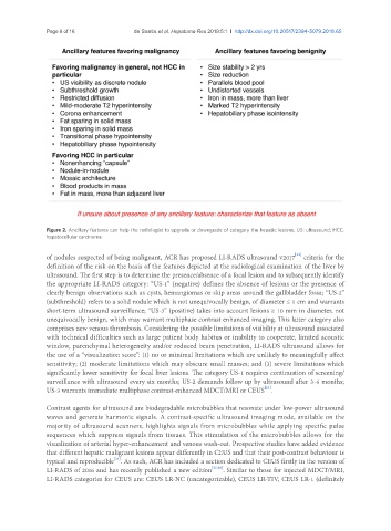
Figure 2. Ancillary features can help the radiologist to upgrade or downgrade of category the hepatic lesions. US: ultrasound; HCC:
hepatocellular carcinoma
[23]
of nodules suspected of being malignant, ACR has proposed LI-RADS ultrasound v2017 criteria for the
definition of the risk on the basis of the features depicted at the radiological examination of the liver by
ultrasound. The first step is to determine the presence/absence of a focal lesion and to subsequently identify
the appropriate LI-RADS category: “US-1” (negative) defines the absence of lesions or the presence of
clearly benign observations such as cysts, hemangiomas or skip areas around the gallbladder fossa; “US-2”
(subthreshold) refers to a solid nodule which is not unequivocally benign, of diameter ≤ 1 cm and warrants
short-term ultrasound surveillance; “US-3” (positive) takes into account lesions ≥ 10 mm in diameter, not
unequivocally benign, which may warrant multiphase contrast enhanced imaging. This latter category also
comprises new venous thrombosis. Considering the possible limitations of visibility at ultrasound associated
with technical difficulties such as large patient body habitus or inability to cooperate, limited acoustic
window, parenchymal heterogeneity and/or reduced beam penetration, LI-RADS ultrasound allows for
the use of a “visualization score”: (1) no or minimal limitations which are unlikely to meaningfully affect
sensitivity; (2) moderate limitations which may obscure small masses; and (3) severe limitations which
significantly lower sensitivity for focal liver lesions. The category US-1 requires continuation of screening/
surveillance with ultrasound every six months; US-2 demands follow up by ultrasound after 3-6 months;
[23]
US-3 warrants immediate multiphase contrast-enhanced MDCT/MRI or CEUS .
Contrast agents for ultrasound are biodegradable microbubbles that resonate under low-power ultrasound
waves and generate harmonic signals. A contrast-specific ultrasound imaging mode, available on the
majority of ultrasound scanners, highlights signals from microbubbles while applying specific pulse
sequences which suppress signals from tissues. This stimulation of the microbubbles allows for the
visualization of arterial hyper-enhancement and venous wash-out. Prospective studies have added evidence
that different hepatic malignant lesions appear differently in CEUS and that their post-contrast behaviour is
[24]
typical and reproducible . As such, ACR has included a section dedicated to CEUS firstly in the version of
LI-RADS of 2016 and has recently published a new edition [25,26] . Similar to those for injected MDCT/MRI,
LI-RADS categories for CEUS are: CEUS LR-NC (uncategorizable), CEUS LR-TIV, CEUS LR-1 (definitely