
Page 307 - Read Online
P. 307
Kohla et al. Transforming growth factor β1 in HCC
Survival functions
TGF group
1.0
Negative
Positive
Negative-censored
Positive-censored
0.8
0.6
Cum survival 0.4
0.2
0.0
0.00 5.00 10.00 15.00 20.00 25.00 30.00
Survival time
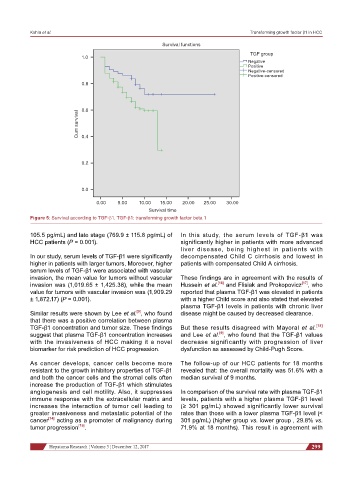
Figure 5: Survival according to TGF-b1. TGF-b1: transforming growth factor beta 1
105.5 pg/mL) and late stage (769.9 ± 115.8 pg/mL) of In this study, the serum levels of TGF-β1 was
HCC patients (P = 0.001). significantly higher in patients with more advanced
liver disease, being highest in patients with
In our study, serum levels of TGF-β1 were significantly decompensated Child C cirrhosis and lowest in
higher in patients with larger tumors, Moreover, higher patients with compensated Child A cirrhosis.
serum levels of TGF-β1 were associated with vascular
invasion, the mean value for tumors without vascular These findings are in agreement with the results of
invasion was (1,019.65 ± 1,425.38), while the mean Hussein et al. [16] and Flisiak and Prokopovicz [17] , who
value for tumors with vascular invasion was (1,909.29 reported that plasma TGF-β1 was elevated in patients
± 1,872.17) (P = 0.001). with a higher Child score and also stated that elevated
plasma TGF-β1 levels in patients with chronic liver
Similar results were shown by Lee et al. , who found disease might be caused by decreased clearance.
[9]
that there was a positive correlation between plasma
TGF-β1 concentration and tumor size. These findings But these results disagreed with Mayoral et al. [18]
suggest that plasma TGF-β1 concentration increases and Lee et al. , who found that the TGF-β1 values
[9]
with the invasiveness of HCC making it a novel decrease significantly with progression of liver
biomarker for risk prediction of HCC progression. dysfunction as assessed by Child-Pugh Score.
As cancer develops, cancer cells become more The follow-up of our HCC patients for 18 months
resistant to the growth inhibitory properties of TGF-β1 revealed that: the overall mortality was 51.6% with a
and both the cancer cells and the stromal cells often median survival of 9 months.
increase the production of TGF-β1 which stimulates
angiogenesis and cell motility. Also, it suppresses In comparison of the survival rate with plasma TGF-β1
immune response with the extracellular matrix and levels, patients with a higher plasma TGF-β1 level
increases the interaction of tumor cell leading to (≥ 301 pg/mL) showed significantly lower survival
greater invasiveness and metastatic potential of the rates than those with a lower plasma TGF-β1 level (<
cancer [14] acting as a promoter of malignancy during 301 pg/mL) (higher group vs. lower group , 29.8% vs.
tumor progression [15] . 71.9% at 18 months). This result in agreement with
Hepatoma Research ¦ Volume 3 ¦ December 12, 2017 299