
Page 303 - Read Online
P. 303
Kohla et al. Transforming growth factor β1 in HCC
appropriate treatment remain the best strategy for 180
reducing mortality. subjects
Transforming growth factor β (TGF-β) superfamily Control Cirrhotic
is known to be involved in embryonic development, group CC group group
adult tissue homeostasis, and disease pathogenesis. (n = 30) (n = 120) (n = 30)
Specifically, it has been shown to control proliferation,
differentiation, apoptosis, migration, extracellular matrix BCLC 0&A BCLC B BCLC C BCLC D
remodeling, immune functions, and tumor invasion/ (n = 30) (n = 30) (n = 30) (n = 30)
[4]
metastasis . TGF-β enhances hepatic stellate cell
activation, stimulates collagen gene transcription and
suppresses matrix metalloproteinases expression.
Thus, TGF-β, as well as its intracellular mediators;
Smad proteins, can be potential therapeutic targets for Serum TGF-b1 level measurement and
liver fibrosis. TGF-β inhibits hepatocyte proliferation, follow-up for 18 months
but it also promotes HCC. TGF-β has been shown to
play both tumor-suppressive at early stage and tumor-
[5]
promoting roles at later stage . At the early stage of
tumorogenesis, TGF-β1 inhibited normal cell growth
Alive
and tumorogenesis by suppressing G1/S phase Deceased (n = 118)
(n = 62)
[6]
transition , in later stages; malignant cells become
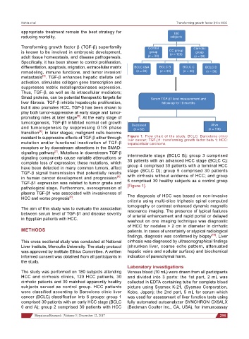
resistant to suppressive effects of TGF-β either through Figure 1: Flow chart of the study. BCLC: Barcelona clinic
mutation and/or functional inactivation of TGF-β liver cancer; TGF-b1: transforming growth factor beta 1; HCC:
hepatocellular carcinoma
receptors or by downstream alterations in the SMAD-
[7]
signaling pathway . Mutations in downstream TGF-β
signaling components cause variable attenuations or intermediate stage (BCLC B); group 3 comprised
complete loss of expression; these mutations, which 30 patients with an advanced HCC stage (BCLC C);
have been detected in many common tumors, affect group 4 comprised 30 patients with a terminal HCC
TGF-β signal transmission that potentially results stage (BCLC D); group 5 comprised 30 patients
[8]
in human cancer development and progression . with cirrhosis without evidence of HCC; and group
TGF-β1 expression was related to tumor grade and 6 comprised 30 healthy subjects as a control group
pathological stage. Furthermore, overexpression of [Figure 1].
plasma TGF-β1 was associated with invasiveness of The diagnosis of HCC was based on non-invasive
[9]
HCC and worse prognosis .
criteria using multi-slice triphasic spiral computed
tomography or contrast enhanced dynamic magnetic
The aim of this study was to evaluate the association resonance imaging. The presence of typical features
between serum level of TGF-β1 and disease severity of arterial enhancement and rapid portal or delayed
in Egyptian patients with HCC. washout on one imaging technique was diagnostic
of HCC for nodules > 2 cm in diameter in cirrhotic
METHODS patients. In cases of uncertainty or atypical radiological
findings, diagnosis was confirmed by biopsy [10] . Liver
This cross sectional study was conducted at National cirrhosis was diagnosed by ultrasonographical findings
Liver Institute, Menoufia University. The study protocol (shrunken liver, coarse echo pattern, attenuated
was approved by institute Ethics Committee. A written hepatic veins and nodular surface) and biochemical
informed consent was obtained from all participants in indication of parenchymal harm.
the study.
Laboratory investigations
The study was performed on 180 subjects attending Venous blood (10 mL) were drawn from all participants
HCC and cirrhosis clinics, 120 HCC patients, 30 and divided into 3 parts: the 1st part, 2 mL was
cirrhotic patients and 30 matched apparently healthy collected in EDTA containing tube for complete blood
subjects served as control group. HCC patients picture using Sysmex K-21, (Sysmex Corporation,
were classified according to Barcelona clinic liver Kobe, Japan); the 2nd part, 5 mL for serum which
cancer (BCLC) classification into 6 groups: group 1 was used for assessment of liver function tests using
comprised 30 patients with an early HCC stage (BCLC fully automated autoanalyzer SYNCHRON CX9ALX
0 and A); group 2 comprised 30 patients with HCC (Beckman Coulter Inc., CA, USA), for immunoassay
Hepatoma Research ¦ Volume 3 ¦ December 12, 2017 295