
Page 151 - Read Online
P. 151
Stambo et al. LC Bead embolization of hepatic neoplasms
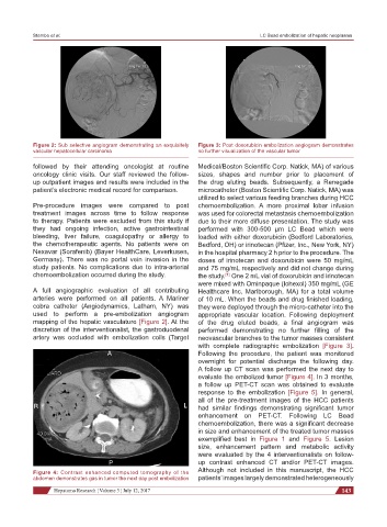
Figure 2: Sub selective angiogram demonstrating an exquisitely Figure 3: Post doxorubicin embolization angiogram demonstrates
vascular hepatocellular carcinoma no further visualization of the vascular tumor
followed by their attending oncologist at routine Medical/Boston Scientific Corp. Natick, MA) of various
oncology clinic visits. Our staff reviewed the follow- sizes, shapes and number prior to placement of
up outpatient images and results were included in the the drug eluting beads. Subsequently, a Renegade
patient’s electronic medical record for comparison. microcatheter (Boston Scientific Corp. Natick, MA) was
utilized to select various feeding branches during HCC
Pre-procedure images were compared to post chemoembolization. A more proximal lobar infusion
treatment images across time to follow response was used for colorectal metastasis chemoembolization
to therapy. Patients were excluded from this study if due to their more diffuse presentation. The study was
they had ongoing infection, active gastrointestinal performed with 300-500 µm LC Bead which were
bleeding, liver failure, coagulopathy or allergy to loaded with either doxorubicin (Bedford Laboratories,
the chemotherapeutic agents. No patients were on Bedford, OH) or irinotecan (Pfizer, Inc., New York, NY)
Nexavar (Sorafenib) (Bayer HealthCare, Leverkusen, in the hospital pharmacy 2 h prior to the procedure. The
Germany). There was no portal vein invasion in the doses of irinotecan and doxorubicin were 50 mg/mL
study patients. No complications due to intra-arterial and 75 mg/mL respectively and did not change during
chemoembolization occurred during the study. the study. One 2 mL vial of doxorubicin and irinotecan
[1]
were mixed with Ominpaque (Iohexol) 350 mg/mL (GE
A full angiographic evaluation of all contributing Healthcare Inc, Marlborough, MA) for a total volume
arteries were performed on all patients. A Mariner of 10 mL. When the beads and drug finished loading,
cobra catheter (Angiodynamics, Latham, NY) was they were deployed through the micro-catheter into the
used to perform a pre-embolization angiogram appropriate vascular location. Following deployment
mapping of the hepatic vasculature [Figure 2]. At the of the drug eluted beads, a final angiogram was
discretion of the interventionalist, the gastroduodenal performed demonstrating no further filling of the
artery was occluded with embolization coils (Target neovascular branches to the tumor masses consistent
with complete radiographic embolization [Figure 3].
A Following the procedure, the patient was monitored
overnight for potential discharge the following day.
A follow up CT scan was performed the next day to
evaluate the embolized tumor [Figure 4]. In 3 months,
a follow up PET-CT scan was obtained to evaluate
response to the embolization [Figure 5]. In general,
all of the pre-treatment images of the HCC patients
R L had similar findings demonstrating significant tumor
enhancement on PET-CT. Following LC Bead
chemoembolization, there was a significant decrease
in size and enhancement of the treated tumor masses
exemplified best in Figure 1 and Figure 5. Lesion
size, enhancement pattern and metabolic activity
were evaluated by the 4 interventionalists on follow-
P up contrast enhanced CT and/or PET-CT images.
Figure 4: Contrast enhanced computed tomography of the Although not included in this manuscript, the HCC
abdomen demonstrates gas in tumor the next day post embolization patients’ images largely demonstrated heterogeneously
Hepatoma Research ¦ Volume 3 ¦ July 12, 2017 143