
Page 53 - Read Online
P. 53
three of the wells were washed three times with phosphate- nonaggressive Alexander cells [Figure 1a]. This piece of data
buffered saline while the remaining three were fixed with further confirms a critical role played by Fascin-1 in HCC and
4% paraformaldehyde (PFA). Washed wells were also fixed cancer cell aggressiveness.
with PFA and then cells in all wells were quantified using
crystal violet. Crystal violet was washed using ddH O and Fascin-1 gene silencing leads to downregulation of both
[13]
2
cells were solubilized using acetic acid. Absorbance was migfilin and VASP
measured at 570 nm using Perkin Elmer EnSpire plate reader We then proceeded with knocking down Fascin-1 gene in
(Waltham, MA, USA). Adhesion was presented as the ratio of both HCC cell lines to better understand its function as
the absorbance at 570 nm of adhered cells (washed) divided well as its effect on known ECM-related proteins. As shown
by the absorbance at 570 nm of the total seeded cells (not in Figure 1b, Fascin-1 was successfully silenced in both cell
washed). The data from two independent experiments were lines transfected with Fascin-1 siRNA compared to the cells
analyzed using the Student’s t-test. P < 0.05 was considered transfected with an NSC siRNA (compare lanes 2 and 4 with
statistically significant. lanes 1 and 3).
Statistical analysis As ECM and actin cytoskeleton are fundamental for HCC
Comparison of means using Statgraphics sof tware progression and aggressiveness, we tested the expression
(Statgraphics Company, Warrenton, VA, USA) was used for the of focal adhesion proteins migfilin (also known as Filamin
statistical analysis. t-test was performed, and P < 0.05 was Binding LIM-protein-1) a novel LIM domain-containing protein
[14]
[15]
considered statistically significant. present both at cell-ECM, cell-cell adhesions, and VASP,
a focal adhesion phosphoprotein known to regulate actin
RESULTS polymerization. [16-18] Interestingly, migfilin and VASP interact
with each other and are implicated in cellular adhesion to
ECM as well as migration. [13]
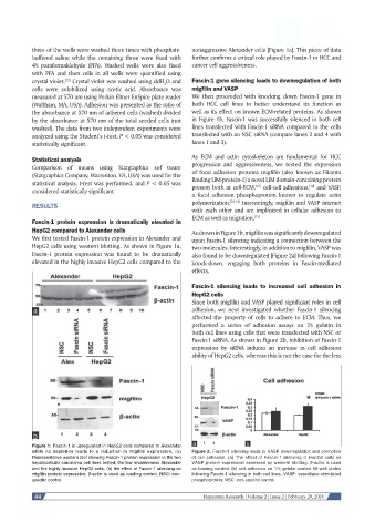
Fascin-1 protein expression is dramatically elevated in
HepG2 compared to Alexander cells As shown in Figure 1b, migfilin was significantly downregulated
We first tested Fascin-1 protein expression in Alexander and upon Fascin-1 silencing indicating a connection between the
HepG2 cells using western blotting. As shown in Figure 1a, two molecules. Interestingly, in addition to migfilin, VASP was
Fascin-1 protein expression was found to be dramatically also found to be downregulated [Figure 2a] following Fascin-1
elevated in the highly invasive HepG2 cells compared to the knock-down, engaging both proteins in Fascin-mediated
effects.
Fascin-1 silencing leads to increased cell adhesion in
HepG2 cells
Since both migfilin and VASP played significant roles in cell
adhesion, we next investigated whether Fascin-1 silencing
affected the property of cells to adhere to ECM. Thus, we
performed a series of adhesion assays on 1% gelatin in
both cell lines using cells that were transfected with NSC or
Fascin-1 siRNA. As shown in Figure 2b, inhibition of Fascin-1
expression by siRNA induces an increase in cell adhesion
ability of HepG2 cells, whereas this is not the case for the less
Figure 1: Fascin-1 is upregulated in HepG2 cells compared to Alexander
while its depletion leads to a reduction in migfilin expression. (a) Figure 2: Fascin-1 silencing leads to VASP downregulation and promotion
Representative western blot showing Fascin-1 protein expression in the two of cell adhesion. (a) The effect of Fascin-1 silencing in HepG2 cells on
hepatocellular carcinoma cell lines tested; the low invasiveness Alexander VASP protein expression assessed by western blotting. β-actin is used
and the highly invasive HepG2 cells; (b) the effect of Fascin-1 silencing on as loading control; (b) cell adhesion on 1% gelatin-coated 96-well plates
migfilin protein expression. β-actin is used as loading control. NSC: non- following Fascin-1 silencing in both cell lines. VASP: vasodilator-stimulated
specific control phosphoprotein; NSC: non-specific control
44 Hepatoma Research | Volume 2 | Issue 2 | February 29, 2016