
Page 58 - Read Online
P. 58
Follow-up and endpoint liver failure. Other complications were listed on Table 2.
Two days af ter RFA, contrast-enhanced computer Further analyses showed that there was no significant
tomography (CT) or magnetic resonance imaging (MRI) difference between the “one-off ” group and other
was performed. If any irregular contrast enhancement treatment groups.
was found inside or beside the ablation zone, additional
RFA would be performed in 1 week. Thirty days after “One-off” ablation and predictive factors for its
the first RFA, contrast-enhanced CT or MRI was carried success
out again. If the enhancing tissue at the tumor site During the CT evaluation 2 days after RFA, there were 416
[6]
disappeared, it was classified as “complete necrosis”. (90.0%) patients who had achieved “complete necrosis”,
Laboratory test of AFP was also used to evaluate the while 46 (10.0%) patients had not. When evaluated at
efficiency of RFA in patients with high pre-operative AFP 6 months after the treatment, 281 (60.8%) patients
levels. Then, patients were regularly followed up in the achieved “one-off ” ablation, while 181 (39.2%) patients
outpatient clinic every 3 months for the first 2 years. In failed. Clinical data were compared between patients
our study, the endpoint was “one-off ” ablation, which who achieved “one- off ” ablation and those who failed
was assessed at the 6th month after RFA.
Table 1: Baseline characteristics of all 462 patients
Statistical analysis Variables n = 462
Data were analyzed with the SPSS statistical software Gender (male/female) (%) 373 (80.7)/89 (19.3)
(SPSS version 20.0, Chicago, IL, USA). Homogeneity Age (years) 56.6 ± 11.0
9
of continuous data was performed by the Gaussianity PLT (×10 /L) 131.1 ± 57.1
12.3 ± 0.95
PT (s)
test, and described as means ± standard deviations or Total bilirubin (µmol/L) 17.2 ± 10.9
median (range) and compared using the unpaired t-test. ALT (IU/L) 86.5 (9.4, 546.8)
Categorical variables were compared using Chi-square test Albumin (g/L) 41.3 ± 4.0
or the Fisher’s exact test, where appropriate. Variables Prealbumin (mg/dL) 186.6 ± 52.1
26.5 (0.6, 584.0)
AFP (ng/mL)
with a P < 0.05 in the univariate analysis would be added Child-Pugh classification
to the multivariate model. In the multivariate analysis, a Class A 442
multiple logistic regression was used to determine the Class B 20
predictors of the success of “one-off ” ablation. Hepatitis background 333
HBV
HCV 7
RESULTS HBV-HCV # 4
HBsAg
Present 333
Baseline data Absent 129
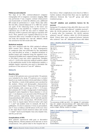
A total of 983 patients were screened while 735 patients HBeAg
were included in the study, 273 patients were excluded Present 117
based on our study exclusion criteria and failure to follow Absent 345
-up. Therefore, a total of 462 patients were enrolled for Tumor size (cm) 2.6 ± 1.1
Tumor location
the analysis. Clinical and demographic characteristics Parenchyma 307
were summarized in Table 1. There were 373 male patients Sub-capsular 155
and 89 females, with a mean age of 56.6 ± 11.0 years. Close to organs 48
Gallbladder
Most patients (85.7%) had a background of viral hepatitis Stomach 22
(hepatitis B and/or hepatitis C). Tumor diameter ≤ 3.0 cm Jejunum 23
and > 3 cm diameter were present in 362 (70.6%) and 136 Pericardium 8
Kidney
(29.4%) patients, respectively. Tumor location included Close to main blood vessels 17
deep-parenchyma (307 patients, 66.5%) and sub-capsular Yes 40
(155 patients, 43.5%). Among them, 109 (23.6%) tumors No 422
were close to organs (space between tumor and organ # Co-occurrence of HBV and HCV. PLT: platelet; PT: prothrombin
< 1 cm) (22 nodules close to stomach, 48 close to time; ALT: alanine aminotransferase; AFP: alpha fetal protein;
[13]
gallbladder, 23 close to jejunum, 8 close to pericardium, HBV: hepatitis B virus; HCV: hepatitis C virus; HBsAg: hepatitis B
and 17 close to kidney), and 40 tumors (3.9%) were close surface antigen; HBeAg: hepatitis B e antigen
to the main blood vessels (between tumor and vessels Table 2: Complications of radiofrequency ablation
< 5 mm) such as post-hepatic vena cava, hepatic vein, Complications Number
[11]
and the portal vein. Severe pain 3
Cholecystitis 6
Complications of RFA Bile leakage 2
1
Intestinal leakage
Most patients experienced mild pain or discomfort Abdominal bleeding 2
during ablation. Twenty patients (4.3%) had one or more Liver abscess 2
complications. One patient died in the hospital due to Pleural effusion 3
Hepatoma Research | Volume 2 | Issue 2 | February 29, 2016 49