
Page 203 - Read Online
P. 203
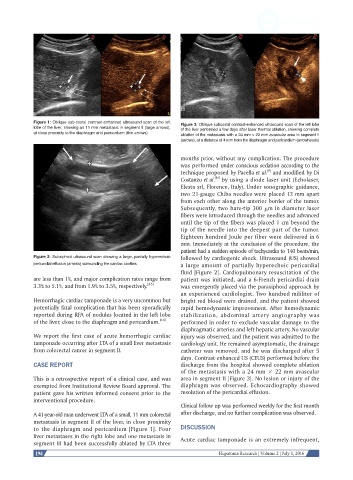
Figure 1: Oblique sub-costal contrast-enhanced ultrasound scan of the left   Figure 3: Oblique subcostal contrast-enhanced ultrasound scan of the left lobe
            lobe of the liver, showing an 11 mm metastasis in segment II (large arrows),   of the liver performed a few days after laser thermal ablation, showing complete
            at close proximity to the diaphragm and pericardium (thin arrows)  ablation of the metastasis with a 24 mm × 22 mm avascular area in segment II
                                                              (arrows), at a distance of 4 mm from the diaphragm and pericardium (arrowheads)

                                                              months prior, without any complication. The procedure
                                                              was performed under conscious sedation according to the
                                                                                           [9]
                                                              technique proposed by Pacella et al.  and modified by Di
                                                                         [10]
                                                              Costanzo et al.  by using a diode laser unit (Echolaser,
                                                              Elesta srl, Florence, Italy). Under sonographic guidance,
                                                              two 21-gauge Chiba needles were placed 12 mm apart
                                                              from each other along the anterior border of the tumor.
                                                              Subsequently, two bare-tip 300 μm in diameter laser
                                                              fibers were introduced through the needles and advanced
                                                              until the tip of the fibers was placed 1 cm beyond the
                                                              tip of the needle into the deepest part of the tumor.
                                                              Eighteen hundred Joule per fiber were delivered in 6
                                                              min. Immediately at the conclusion of the procedure, the
                                                              patient had a sudden episode of tachycardia to 140 beats/min,
            Figure 2: Subxiphoid ultrasound scan showing a large, partially hyperechoic   followed by cardiogenic shock. Ultrasound (US) showed
            pericardial effusion (arrows) surrounding the cardiac cavities  a large amount of partially hyperechoic pericardial
                                                              fluid [Figure 2]. Cardiopulmonary resuscitation of the
            are less than 1%, and major complication rates range from   patient was initiated, and a 6-French pericardial drain
            3.3% to 5.1%, and from 1.9% to 3.5%, respectively. [3-5]  was emergently placed via the paraxiphoid approach by
                                                              an experienced cardiologist. Two hundred mililiter of
            Hemorrhagic cardiac tamponade is a very uncommon but   bright red blood were drained, and the patient showed
            potentially fatal complication that has been sporadically   rapid hemodynamic improvement. After hemodynamic
            reported during RFA of nodules located in the left lobe   stabilization,  abdominal  artery  angiography  was
            of the liver, close to the diaphragm and pericardium. [6-8]  performed in order to exclude vascular damage to the
                                                              diaphragmatic arteries and left hepatic artery. No vascular
            We report the first case of acute hemorrhagic cardiac   injury was observed, and the patient was admitted to the
            tamponade occurring after LTA of a small liver metastasis   cardiology unit. He remained asymptomatic, the drainage
            from colorectal cancer in segment II.             catheter was removed, and he was discharged after 5
                                                              days. Contrast-enhanced US (CEUS) performed before the
            CASE REPORT                                       discharge from the hospital showed complete ablation
                                                              of the metastasis with a 24 mm × 22 mm avascular
            This is a retrospective report of a clinical case, and was   area in segment II [Figure 3]. No lesion or injury of the
            exempted from Institutional Review Board approval. The   diaphragm was observed. Echocardiography showed
            patient gave his written informed consent prior to the   resolution of the pericardial effusion.
            interventional procedure.
                                                              Clinical follow up was performed weekly for the first month
            A 41-year-old man underwent LTA of a small, 11 mm colorectal   after discharge, and no further complication was observed.
            metastasis in segment II of the liver, in close proximity
            to the diaphragm and pericardium [Figure 1]. Four   DISCUSSION
            liver metastases in the right lobe and one metastasis in   Acute cardiac tamponade is an extremely infrequent,
            segment III had been successfully ablated by LTA three
             194                                                           Hepatoma Research | Volume 2 | July 1, 2016