
Page 106 - Read Online
P. 106
inhibitor and 1 mmol/L PMSF. The protein concentration Statistical analysis
was determined by Bradford assay (Bio-Rad, Hercules, Statistical analyses were carried out by unpaired or paired
CA, USA). Equal amounts of protein were loaded and t-test as appropriate. All data are reported as mean ± standard
separated by sodium dodecyl sulfate-polyacrylamide gel deviation. P < 0.05 were considered significant.
electrophoresis, and the proteins were transferred on
to a PVDF membrane (Millipore, Billerica, MA, USA). For RESULTS
Western blotting, the membranes were incubated with
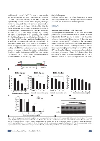
anti-actin (Sigma, Steinheim, Germany), anti-HBx (Chemicon, Sorafenib suppresses HBV gene expression
Danvers, MA, USA), anti-flag (Cell Signaling, Beverly, To investigate the anti-viral effect of sorafenib, we obtained
MA, USA), anti-SAPK/JNK (Cell Signaling), anti-p-SAPK/ promoters of genes contained in the HBV genome. As shown
JNK (Cell Signaling), anti-c-jun (Santa Cruz Biotechnology, in Figure 1a, the HBV genome contains 4 promoters and 2
Santa Cruz, CA, USA), anti-p-c-jun (Santa Cruz Biotechnology) enhancers that regulate HBV replication. Of these, the most
or anti-FXR (Santa Cruz Biotechnology) antibodies in important one during HBV replication is the precore/core
tris-buffered saline with Tween 20 (TBST) containing 1% promoter, which has an effect on transcription of pregenomic
Tween 20 supplemented with 3% nonfat dried milk. After RNA from cccDNA. The ×1.3 HBV-Cp-luc construct contains
[5]
washing with TBST, the blotted membranes were incubated the core promoter [Figure 1a]. The promoter activities of the
with the peroxidase-conjugated secondary antibody (Santa HBV core, X, and preS1 genes were decreased by sorafenib in
Cruz Biotechnology). After washing TBST, the proteins were a dose-dependent manner [Figure 1b-d]. To investigate if the
visualized by the ECL development reagent (Amersham decrease of promoter activities by sorafenib was induced by
Pharmacia Biotech, Piscataway, NJ, USA). cell death, a cell viability assay was performed. The results
a
b c d e
f g
Figure 1: Sorafenib suppresses hepatitis B virus (HBV) gene expression. (a) Structure of the ×1.3 HBV-luc plasmid; (b-d) the effects of sorafenib on HBV promoter
activity. HepG2 or Chang cells were transfected with ×1.3 HBV-luc, pGL4-X or pGL4-preS1 constructs for 24 h and treated with sorafenib. The cell lysates were
analyzed for luciferase activity. *P < 0.05, **P < 0.01, compared with the indicated cells; (e) cell viability assay; (f and g) the effect of sorafenib on HBV RNA or
protein levels. HepG2 cells were transfected with 1.2 mer HBV construct for 24 h and treated with sorafenib. The indicated mRNA levels were detected by real-time
polymerase chain reaction. Total proteins were prepared from the cells, and then HBx protein levels were detected by Western blotting
Hepatoma Research | Volume 1 | Issue 2 | July 15, 2015 99