
Page 102 - Read Online
P. 102
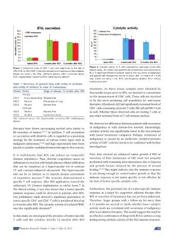
Figure 2: Cytolytic activity of T cells measured by adenylate kinase (AK)
                                 +
          Figure 1: Stimulation index of CD8  T cells was determined by the ratio of   release assay. AK release was quantifi ed by luminescence and expressed as
                      −
             +
                +
                         +
          IFNγ CD8  vs. IFNγ CD8  T cells after stimulation with autologous tumor antigens.   RLU. A signifi cant difference between patients with recurrence of malignancy
          Shown are mean ± SD. IFNγ: interferon gamma; CRC: colorectal cancer;   and patients with disease-free survival is shown after 12 months (P < 0.05)
          HCC: hepatocellular carcinoma; RFA: radiofrequency ablation
                                                              only. Shown are mean ± SD.   RFA: radiofrequency ablation; RLU: relative
                                                              luminescence units
          Table 1: Summary of general data with entity of cirrhosis
          and entity of primary in case of metastases
                                                              treatment. As these tissue samples were obtained by
          Patient Entity       Stage of disease 12 months after RFA
                                                              fine-needle biopsy prior to RFA, we decided to concentrate
                Primary
                                                              on the measurement of CD8  cells. These cells are reported
                                                                                     +
          CRC1    Colon descending Disease free
          CRC2    Rectum       Metastases to lung             to be the most promising cell population for anti-tumor
          CRC3    Rectum       Disease free                   therapies. All patients still had significantly increased levels of
                                                                  +
                Cirrhosis                                     CD8  cells containing cytotoxic T cells, NK cell and NK T cells
          HCC1    Alcohol      Disease free                   as well. Whether these observed cells are resting T cells or
          HCC2    Alcohol      Local recurrence               any other activated form of T cell remains unclear.
          CRC: colorectal cancer; HCC: hepatocellular carcinoma; RFA: radiofrequency
          ablation
                                                              We observed no differences between patients with recurrence
          therapies have shown encouraging survival rates similar to   of malignancy or with disease-free survival. Interestingly,
          R0 resection of tumors. [1,2,11]  In addition, T cell vaccination   cytolytic activity was significantly lower in the two patients
          or vaccination with dendritic cells is regarded as a promising   with tumor recurrence compared. Perhaps, recurrence of
          strategy for the treatment of various tumor types such as   malignancy is caused by an ineffective cytolytic/cytotoxic
                                                                          +
          malignant melanomas, [12-20]  and high expectations have been   activity of CD8  cells but needs to be confirmed with further
          placed on cytokine-modulated immunotherapy for liver tumors.  investigations.
          It is well-known that RFA can induce an unspecific   Prior data showed an enhanced tumor growth if RFA or
          immune stimulation. Thus, thermal coagulation causes an   resection of liver metastases of CRC were not properly
          inflammatory reaction with lymph-plasma-cellular infiltration   performed with remaining micrometastases due to hypoxia
          that can be visualized as a hypervascular rim in contrast   and growth factors induced by the process of wound
          CT and contrast-enhanced ultrasound. This hypervascular   healing. [21,22]  This might indicate that the immune response
          rim can be so intense as to impede proper assessment   is not strong enough to control tumor growth or that the
                              [5]
          of treatment success.  We recently demonstrated a   immune response is not tumor specific or not effective by
          specific T cell response, after RFA application, toward the   the lack of tumor-specific cytolytic cells.
                                                       [6]
          orthotopic VX 2-tumor implantation in rabbit livers.  In
          the clinical setting, it was also shown that a tumor-specific   Furthermore, the potential role of a tumor-specific immune
          immune response could be detected in patients after RFA.   response as a target for supportive adjuvant therapy after
          Our preliminary study showed a significant appearance of   RFA or resection of metastases has not yet been addressed.
          tumor-specific CD4  and CD8  T cells in peripheral blood up   Therefore, larger groups with a follow-up for more than
                          +
                                  +
          to 8 weeks after RFA. The cytolytic activity of isolated PBMC   6-12 months are needed to clarify whether lower cytolytic
          was also significantly elevated. [8]                activity levels are correlated with recurrence of malignancy
                                                              after local ablative therapies. This would suggest the need for
          In this study, we investigated the presence of tumor-specific   an effective combination of drugs with RFA to achieve a long
          T cells and the cytolytic activity 12 months after RFA   lasting strong cytolytic activity of the Th2 immune response.


               Hepatoma Research | Volume 1 | Issue 2 | July 15, 2015                                        95