
Page 100 - Read Online
P. 100
Thiruchelvam et al. Hepatoma Res 2021;7:22 I http://dx.doi.org/10.20517/2394-5079.2020.144 Page 9 of 18
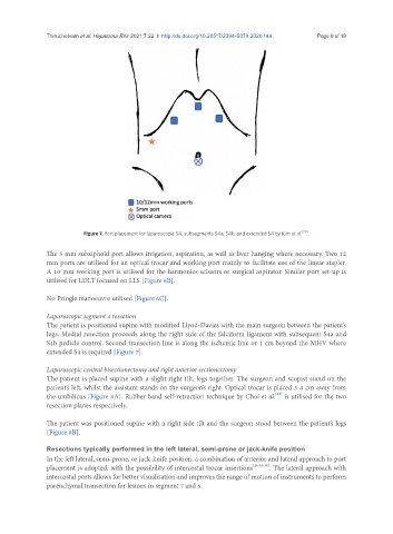
Figure 7. Port placement for laparoscopic S4, subsegments S4a, S4b, and extended S4 by Kim et al. [29] .
The 5 mm subxiphoid port allows irrigation, aspiration, as well as liver hanging where necessary. Two 12
mm ports are utilised for an optical trocar and working port mainly to facilitate use of the linear stapler.
A 10 mm working port is utilised for the harmonics scissors or surgical aspirator. Similar port set-up is
utilised for LDLT focused on LLS [Figure 6B].
No Pringle manoeuvre utilised [Figure 6C].
Laparoscopic segment 4 resection
The patient is positioned supine with modified Llyod-Davies with the main surgeon between the patient’s
legs. Medial resection proceeds along the right side of the falciform ligament with subsequent S4a and
S4b pedicle control. Second transection line is along the ischemic line or 1 cm beyond the MHV where
extended S4 is required [Figure 7].
Laparoscopic central bisectionectomy and right anterior sectionectomy
The patient is placed supine with a slight right tilt, legs together. The surgeon and scopist stand on the
patient’s left, whilst the assistant stands on the surgeon’s right. Optical trocar is placed 3-4 cm away from
[31]
the umbilicus [Figure 8A]. Rubber band self-retraction technique by Choi et al. is utilised for the two
resection planes respectively.
The patient was positioned supine with a right side tilt and the surgeon stood between the patient’s legs
[Figure 8B].
Resections typically performed in the left lateral, semi-prone or jack-knife position
In the left lateral, semi-prone, or jack-knife position, a combination of anterior and lateral approach to port
placement is adopted, with the possibility of intercostal trocar insertions [16,34-38] . The lateral approach with
intercostal ports allows for better visualisation and improves the range of motion of instruments to perform
parenchymal transection for lesions in segment 7 and 8.