
Page 102 - Read Online
P. 102
Thiruchelvam et al. Hepatoma Res 2021;7:22 I http://dx.doi.org/10.20517/2394-5079.2020.144 Page 11 of 18
A B
C
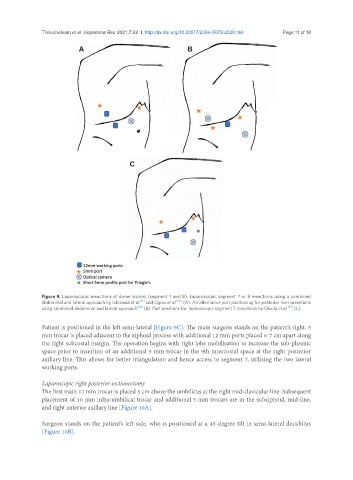
Figure 9. Laparoscopic resections of dome lesions (segment 7 and 8). Laparoscopic segment 7 or 8 resections using a combined
[11]
abdominal and lateral approach by Ishizawa et al. and Ogiso et al. [36] (A). An alternative port positioning for posterior liver resections
using combined abdominal and lateral approach [40] (B). Port positions for laparoscopic segment 7 resections by Okuda et al. [37] (C).
Patient is positioned in the left semi-lateral [Figure 9C]. The main surgeon stands on the patient’s right. 5
mm trocar is placed adjacent to the xiphoid process with additional 12 mm ports placed 6-7 cm apart along
the right subcostal margin. The operation begins with right lobe mobilisation to increase the sub-phrenic
space prior to insertion of an additional 5 mm trocar in the 9th intercostal space at the right posterior
axillary line. This allows for better triangulation and hence access to segment 7, utilising the two lateral
working ports.
Laparoscopic right posterior sectionectomy
The first main 12 mm trocar is placed 5 cm above the umbilicus at the right mid-clavicular line. Subsequent
placement of 10 mm infra-umbilical trocar and additional 5 mm trocars are in the subxiphoid, mid-line,
and right anterior axillary line [Figure 10A].
Surgeon stands on the patient’s left side, who is positioned at a 45-degree tilt in semi-lateral decubitus
[Figure 10B].