
Page 101 - Read Online
P. 101
Page 10 of 18 Thiruchelvam et al. Hepatoma Res 2021;7:22 I http://dx.doi.org/10.20517/2394-5079.2020.144
A B
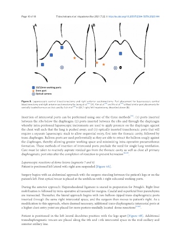
Figure 8. Laparoscopic central bisectionectomy and right anterior sectionectomy. Port placement for laparoscopic central
bisectionectomy and right anterior sectionectomy by Jeung et al. [30] (A). Kim et al. [32] and Ho et al. [33] utilised similar port placements for
centrally located tumours as that used by Suh et al. [22] in LDLT right/left hepatectomy, described above (B).
[35]
Insertion of intercostal ports can be performed using one of the three methods : (1) ports inserted
between the ribs below the diaphragm; (2) ports inserted between the ribs and through the diaphragm
whereby intra-peritoneal laparoscopic instruments are used to apply pressure on the diaphragm against
the chest wall such that the lung is pushed away; and (3) optically inserted transthoracic ports that will
require a separate laparoscopic stack to allow sequential entry, first into the thoracic cavity, followed by
trans-diaphragm. Balloon-ports are used preferentially as they are able to retract the balloon snugly against
the diaphragm, thereby allowing greater working space and minimizing intra-operative pneumothorax
formation. These methods of insertion of intercostal ports preclude the need for single lung ventilation.
Care must be taken to routinely aspirate residual gas from the thoracic cavity as well as close all primary
diaphragmatic port sites after the completion of resection to prevent herniation [36,39] .
Laparoscopic resections of dome lesions (segments 7 and 8)
Patient is positioned left lateral with right arm suspended [Figure 9A].
Surgery begins with an abdominal approach with the surgeon standing between the patient’s legs or on the
patient’s left. First optical trocar is placed at the umbilicus with 3 right subcostal working ports.
During the anterior approach: Hepatoduodenal ligament is snared in preparation for Pringle’s. Right liver
mobilisation is followed by intra-operative ultrasound for margins. Caudal and superficial liver parenchyma
are transected. Thereafter, the lateral approach begins with two balloon-tipped trans-diaphragmatic ports
inserted through the same right intercostal space, and the surgeon then moves to patient’s right. As a
modification to this approach, where deemed necessary, additional trans-diaphragmatic intercostal ports at
a higher chest entry point are placed for more postero-medially located dome resections [35,40] .
Patient is positioned in the left lateral decubitus position with the legs apart [Figure 9B]. Additional
transdiaphragmatic trocars are placed along the 9th and 11th intercostal space in the mid-axillary and
anterior axillary line.