
Page 104 - Read Online
P. 104
Thiruchelvam et al. Hepatoma Res 2021;7:22 I http://dx.doi.org/10.20517/2394-5079.2020.144 Page 13 of 18
A B
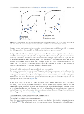
Figure 11. Right-sided resections performed in the semi-prone position. Port positioning by Ikeda et al. [16] for right sided resections in the
semi-prone position (A). Laparoscopic right posterior sectionectomy in the left semi-prone position with jack-knife (B).
the right hepatic vein improves as the transection proceeds in a caudal-cranial fashion, with the remnant
[45]
liver falling away from the resected liver, which remains fixed to the retroperitoneum .
This gravitational shift may not be so apparent in cases where the patient is positioned in a left semi-
lateral decubitus or supine with right-sided 30-degree tilt. In these instances, the right lobe of the liver is
often fully mobilised to allow for not only the visualisation of the right hepatic vein at its origin, but also
[46]
to facilitate a better view of the resection plane . Full mobilisation allows retraction of the liver antero-
medially, such that the resection plane along the right hepatic vein shifts more medially and sits in a
vertical axis. Often stay-sutures or rubber-band retraction technique can be utilised on both sides of the
resection line to splay open the resection plane and enhance exposure.
Similar right-sided resections performed instead in the semi-prone position
The surgeon stands on the left cranial side of the patient with the camera assistant on the left caudal side
of patient. The first port is placed in the right para-rectal line, 10 cm below the costal margin. Additional 3
working ports (5 or 12 mm) are placed in the right pararectal, anterior axillary, and posterior axillary line.
An intercostal port in the 7th intercostal space in the anterior axillary line is inserted for segment 7, 8, and
right superior caudate resections [Figure 11A].
A total of 5-6 trocars are utilised in 2 rows. The optical camera utilised in this series is a 5 mm camera
positioned 5 cm to the right and above the umbilicus, with an additional 12 mm trocar placed at the right
pararectal line at the same level. An upper row of 5 mm trocars is placed along the right subcostal margin
in the right mid-axillary and mid-clavicular line, with an additional 12 mm sub-xiphoid trocar. Dissection
similarly begins with hilar inflow control of the posterior sectoral and thereafter full right lobe mobilisation
followed by parenchyma transection [Figure 11B].
LESS COMMONLY EMPLOYED SURGICAL APPROACHES
Trans-diaphragmatic thoracoscopic approach
Trans-thoracic trans-diaphragmatic surgical approach has also been described for lesions in the dome
of the liver [12,47-49] . This technique involves port placements into the thoracic cavity followed by trans-