
Page 88 - Read Online
P. 88
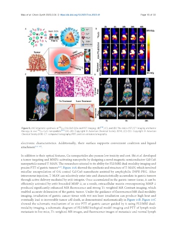
Mao et al. Chem Synth 2023;3:26  https://dx.doi.org/10.20517/cs.2022.41         Page 15 of 33


                                         64
                Figure 9. (A) Schematic synthesis of  Cu-CIS/ZnS QDs and PET imaging; (B) [119] , (C), and (D) The micro-PET/CT imaging and tumor
                          64
                therapy in vivo  Cu-CuS  nanoparticle [120] . (A)-(B): Copyright © American Chemical Society 2014; (C)-(D): Copyright © American
                Chemical Society 2010. CT: computed tomography; PET: positron emission tomography.
               electronic  characteristics.  Additionally,  their  surface  supports  convenient  coalition  and  ligand
               attachment [123-125] .


               In addition to their optical features, Cu nanoparticles also possess low toxicity and cost. Shi et al. developed
               a tumor-targeting and MMP2-activating nanoprobe by designing a novel magnetic semiconductor Gd/CuS
               nanoparticle named T-MAN. The researchers attested to its ability for FLI/MRI dual-modality imaging and
               precise PTT of gastric tumors . Figure 10A showed the synthesis and structure of T-MAN, which involved
                                        [126]
               micellar encapsulation of OA-coated Gd/CuS nanosheets assisted by amphiphilic DSPE-PEG. After
               intravenous injection, T-MAN can selectively enter into and characteristically accumulate in gastric tumors
               through active delivery mediated by αvβ3 integrin. Once accumulated in the gastric tumor tissue, it can be
               efficiently activated by αvβ3-bounded MMP-2; as a result, extracellular matrix-overexpressing MMP-2
               produced significantly enhanced NIR fluorescence and strong T1-weighted MR Contrast imaging, which
               enabled accurate delineation of the gastric tumor. Under the guidance of fluorescence/MR dual-modality
               imaging, irradiation of gastric cancer tissue with 808 nm laser irradiation can produce high heat and
               eventually lead to irreversible tumor cell death, as demonstrated mathematically in Figure 10B. Figure 10C
               showed the schematic mechanism of in vivo PTT of gastric cancer guided by it using FLI/MRI dual-
               modality imaging, a schematic diagram of FLI/MRI biological model imaging and PTT of lymph node
               metastasis in live mice, T1-weighted MR images, and fluorescence images of metastatic and normal lymph