
Page 85 - Read Online
P. 85
Page 12 of 33 Mao et al. Chem Synth 2023;3:26 https://dx.doi.org/10.20517/cs.2022.41
3+
3+
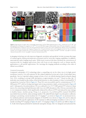
Figure 7. Applications of copper sulfur compounds in biosensing. (A) The TEM characterization of mCu S@SiO @Y O :Yb /Er and
2
2-x
3
2
the application of fingerprint recognition in (B) [103] ; (C) and (D) TEM images of hydrophobic Cu S NPs and amphiphilic Cu S NCs
7 4
7 4
included fabrication strategy for Cu S NCs and photothermal effects of Cu S NCs under a given power density versus the irradiation
7 4
7 4
time, and scheme for photothermal imaging of fingerprints. (The photothermal tests were conducted on the thin film of Cu S NC
7 4
powder with excitation at 808 nm); (E) the photothermal latent fingerprints imaging method [104] . (A)-(B): Copyright © American
Chemical Society 2017; (C)-(E): Copyright © American Chemical Society 2015.
in imaging technology not only improves diagnostic sensitivity and specificity but also increases the range
of contrast agents, making nanomaterials a dominant tool for molecular imaging capabilities. In addition,
nanomaterials make imaging much easier. While many recent articles have discussed the conveniences of
nanomaterials for imaging applications, they only focus on sub-categories such as disease-specific
applications or cell-specific applications. We rearrange the imaging methods according to the order of
clinical use as follows.
Computed tomography
Computed tomography (CT) technology plays a compelling role in the clinic due to its high spatial
resolution; however, it is still restricted by the related radiation burden and a lack of individual tissue
specificity. Cui et al. reported a plain strategy to form a Cu S -Au hybrid structure based on heavily doped
7 4
Cu S NCs, resulting in strong LSPR absorption at approximately 808 nm . Based on this, the F
[105]
19
7 4
molecules were bonded on the surface of the hybrid structure by click chemistry to form Cu S @Au@PSI- F
19
7 4
nanoparticles (named as CNs) in Figure 8A. These NCs were successfully used for coinstantaneous CT/ F-
19
MRI guided PTT with low background signal and strong penetration depth, as demonstrated in Figure 8B
and C. Subsequently, the polymorphous CNs were utilized for the CT/ F-MRI-guided PTT ablation of deep
19
tumors. By connecting small gold domains with Cu S , the LSPR absorption of the CNs was converted to the
7 4
in vivo transparent window, which significantly enhanced the efficacy of PTT and reduced the laser damage
to healthy tissues. Another group, Wang et al., prepared a Cu Se/Bi Se @PEG (named CB3@PEG)
3
2-x
2
nanohybrid structure by the cation exchange method. The added Bi had a high X-ray absorption coefficient,
making it a promising contrast medium for CT imaging , as shown in Figure 8D. As mentioned above,
[106]
the synthesis pathways and mechanism diagram of CB3@PEG were shown in Figure 8D, and the adjacent
photos showed the specific application of CB3 material in different imaging modalities in vitro or in vivo,
mainly including PTT imaging in Figure 8E and F, CT imaging in Figure 8G and H, and MRI in Figure 8I-
K. It also showed great potential for multimode synergistic therapy. Shi et al. designed and synthesized the