
Page 31 - Read Online
P. 31
Page 982 Lee et al. Cancer Drug Resist 2020;3:980-91 I http://dx.doi.org/10.20517/cdr.2020.73
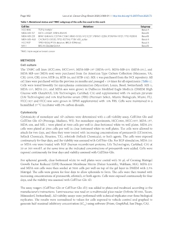
Table 1. Mutational status and TNBC subgroup of the cells line used in this work
Cell line Mutations Subgroup
HCC1806 TCF12-A482V Basal A
MDA-MB-157 FAT4-L4468P; MSH6-R644S Basal B
MDA-MB-231 BRAF-G4646V; CD79A-C106Y; KRAS-G13D; NF2-E231*; PBRM1-I228V; PDGFRA-Y172F; TP53-R280K Basal B
MDA-MB-468 CACNA1D-E953D; TP53-R273H, PTEN V85_splice Basal A
HCC1937 TP53-R308; PTEN deletion; BRCA-5396insC Basal A
MX-1 BRCA1-3363delGAAA Basal B
TNBC: triple negative breast cancer
METHODS
Cell culture
The TNBC cell lines HCC1806, HCC1937, MDA-MB-157 (MDA-157), MDA-MB-231 (MDA-231), and
MDA-MB-468 (MDA-468) were purchased from the American Type Culture Collection (Manassas, VA;
CRL-2335, CRL-2336, HTB-24, HTB-26, and HTB-132). MX-1 was purchased from the NCI repository. All
cell lines were purchased within the previous 24 months and passaged < 15 times for all experiments [Table 1].
Cells were tested biweekly for mycoplasma contamination (MycoAlert, Lonza, Basel, Switzerland). MX-1,
MDA-157, MDA-231, and MDA-468 were grown in Dulbecco Modified Eagle Medium (DMEM High
Glucose with GlutaMAX, Life Technologies, Carlsbad, CA) and supplemented with 1% sodium pyruvate
(Life Technologies) and 10% fetal bovine serum (FBS) (Premium Select, Atlantic Biologicals, Miami, FL).
HCC1937 and HCC1806 were grown in RPMI supplemented with 10% FBS. Cells were maintained in a
humidified 37 °C incubator with 5% carbon dioxide.
Cytotoxicity
Cytotoxicity of monolayer and 3D cultures were determined with a cell viability assay, CellTiter-Glo and
CellTiter-Glo 3D (Promega, Madison, WI). For monolayer experiments, HCC1806, HCC1937, MDA-157,
MDA-468, and MX-1 were plated at 5000 cells per well in clear-bottomed white 96-well plates. MDA-231
cells were plated at 2500 cells per well in clear-bottomed white 96-well plates. The cells were allowed to
attach for two days, and then they were treated with increasing concentrations of prexasertib (LY2606368,
Selleck Chemicals, Houston, TX), erlotinib (Selleck Chemicals), or both agents. The cells were exposed
continuously for four days, and the viability was assessed with CellTiter-Glo. For EGF stimulation, MDA-231
or MDA-468 were treated with EGF (human recombinant proteins, Life Technologies, Carlsbad, CA) at
50 or 500 nmol/L at the same time as the indicated concentrations of prexasertib were added. Cells were
exposed continuously for four days and viability assessed with CellTiter-Glo.
For spheroid growth, clear-bottomed white 96-well plates were coated with 50 µL of Corning Matrigel
Growth Factor Reduced (GFR) Basement Membrane Matrix (Fisher Scientific, Waltham, MA). MDA-231
and MDA-468 cells were then seeded at 5000 cells per well on top of the gel layer in DMEM with 2.5%
Matrigel. The cells were grown for four days to allow spheroids to form. The cells were then treated with
increasing concentrations of prexasertib, erlotinib, or both agents. Cells were exposed continuously for four
days, and the viability was assessed with CellTiter-Glo 3D.
The assay reagent (CellTiter-Glo or CellTiter-Glo 3D) was added to plates and incubated according to the
manufacturer’s instructions. Luminescence was read on a multimodal plate reader (Infinite M1000, Tecan,
Männedorf, Switzerland). All viability assays were performed with technical triplicates over three biological
replicates. The results were normalized to values for cells exposed to vehicle control and graphed to
generate half-maximal inhibitory concentration (IC ) using software (Prism, GraphPad, San Diego, CA).
50