
Page 32 - Read Online
P. 32
Lee et al. Cancer Drug Resist 2020;3:980-91 I http://dx.doi.org/10.20517/cdr.2020.73 Page 983
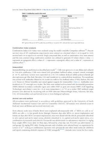
Table 2. Antibodies used in this work
Dilution Reagent Source
1:500 p-EGFR Tyr 1068 Cell Signaling, Danvers, MA
1:1,000 AKT Cell Signaling
BAD Cell Signaling
BRAF Cell Signaling
EGFR Cell Signaling
ERK Cell Signaling
GAPDH Santa Cruz Biotechnology, Dallas, TX
MEK Cell Signaling
p-AKT Ser 473 Cell Signaling
p-BAD Ser 112 Cell Signaling
p-BRAF Ser 445 Cell Signaling
p-ERK Thr 202/204 Cell Signaling
p-MEK Ser 217/221 Cell Signaling
1:30,000 β-actin Life Technologies
AKT: protein kinase B; EGFR: epidermal growth factor receptor; ERK: extracellular signal-regulated kinase
Combination index analysis
Combination Index (CI) values were analyzed using the readily available CompuSyn software . Percent
[20]
survival data of 3D combination experiments were entered as a decimal where 1.00 is equal to 100%
survival and 0.00 is equal to 0% survival. Single compound data, as well as combination data, were entered
and the non-constant drug combination analysis was utilized to generate CI values where a value > 1
represents an antagonist effect, a value of < 1 represents a synergistic effect, and a value of 1 represents an
[20]
additive effect .
Immunoblot
[21]
Immunoblotting was performed as described previously . Cells were grown in 10-cm dishes and cultured
to 70%-80% confluence. Cells were rinsed with phosphate-buffered saline, scraped, stored overnight
at -80 °C, and lysed. Lysates were separated on a 4%-15% sodium dodecyl sulfate-polyacrylamide gel
electrophoresis gel (Bio-Rad, Hercules, CA) and transferred to a nitrocellulose membrane. The membrane
was probed with antibodies diluted in 5% nonfat dry milk in Tris-buffered saline (VWR, Radnor, PA) and
0.1% Tween 20 (Fisher Scientific) and raised against reagents for immunoblot [Table 2]. Antibodies were
incubated at 4 °C overnight on a rocker. The blots were washed and incubated with horseradish peroxidase
(HRP)-labeled secondary antibodies (goat anti-rabbit-HRP or goat anti-mouse-HRP) (Cell Signaling
Technology) and diluted 1:5000 for 1 h at room temperature (~23 °C) on a rocker. HRP antibody target
proteins were detected by incubating with an HRP substrate (WesternBright Sirius, Advansta, San Jose,
CA). All immunoblots were performed in two or more biological replicates.
Animal care and welfare
All procedures were performed in accordance with guidelines approved by the University of South
Alabama’s Institutional Animal Care and Use Committee (IACUC). All animals were allowed access to
food and water ad libitum and received veterinary care.
6
7
Forty athymic nude mice (Charles River) were implanted subcutaneously with 10 MDA-231 cells or 10
MDA-468 cells 1:1 in low growth factor Matrigel. Nineteen days after MDA-231 tumor implantation and
twenty-six days after MDA-468 tumor implantation, mice were dosed with the vehicle, prexasertib (dissolved
in 40% captisol and sterile water) alone, erlotinib (dissolved in 4% captisol and sterile water) alone, or a
combination of prexasertib and erlotinib. Prexasertib was administered via subcutaneous injection [10 mg/kg
body weight (BW)] twice daily for three days, followed by a four-day rest. Erlotinib was administered via
oral gavage (50 mg/kg BW) daily. Dosing amounts and schedules were selected based on experiments by
[8]
Lowery et al. looking at combination exposures with prexasertib and other small molecule inhibitors
and reports examining erlotinib response of TNBC xenografts [8,9,22,23] . Animals were treated for four weeks.