
Page 35 - Read Online
P. 35
Page 986 Lee et al. Cancer Drug Resist 2020;3:980-91 I http://dx.doi.org/10.20517/cdr.2020.73
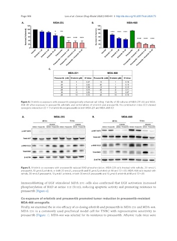
Figure 4. Erlotinib co-exposure with prexasertib synergistically enhanced cell killing. Viability of 3D cultures of MDA-231 (A) and MDA-
468 (B) after exposure to prexasertib, erlotinib, and combinations of erlotinib and prexasertib; the combination index (CI) showed
synergistic interaction (CI < 1) of erlotinib and prexasertib in both MDA-231 and MDA-468 (C)
Figure 5. Erlotinib co-exposure with prexasertib reduced BAD phosphorylation. MDA-231 cells treated with vehicle, 20 nmol/L
prexasertib, 10 µmol/L erlotinib, or both 20 nmol/L prexasertib and 10 µmol/L erlotinib at 48 and 72 h (A); MDA-468 cells treated with
vehicle, 20 nmol/L prexasertib, 10 µmol/L erlotinib, or both 20 nmol/L prexasertib and 10 µmol/L erlotinib at 48 and 72 h (B)
Immunoblotting of EGF stimulated MDA-231 cells also confirmed that EGF activation increased
phosphorylation of BAD at serine 112 (S112), reducing apoptotic activity and promoting resistance to
prexasertib [Figure 6].
Co-exposure of erlotinib and prexasertib promoted tumor reduction in prexasertib-resistant
MDA-468 xenografts
Finally, we examined the in vivo efficacy of co-dosing erlotinib and prexasertib in MDA-231 and MDA-468.
MDA-231 is a commonly used preclinical model cell for TNBC with representative sensitivity to
prexasertib [Figure 1]. MDA-468 was selected for its resistance to prexasertib. Athymic nude mice were