
Page 53 - Read Online
P. 53
Jabbari et al. Art Int Surg. 2025;5:200-9 https://dx.doi.org/10.20517/ais.2024.77 Page 202
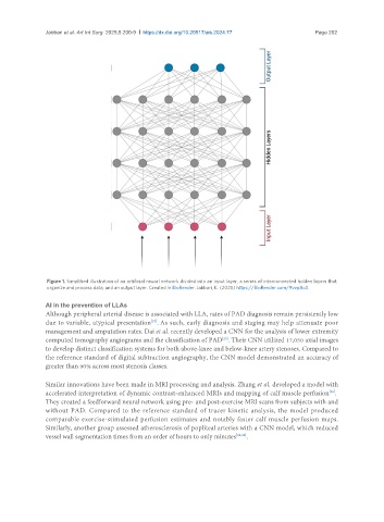
Figure 1. Simplified illustration of an artificial neural network divided into an input layer, a series of interconnected hidden layers that
organize and process data, and an output layer. Created in BioRender. Jabbari, K. (2025) https://BioRender.com/9vvp8o3.
AI in the prevention of LLAs
Although peripheral arterial disease is associated with LLA, rates of PAD diagnosis remain persistently low
[27]
due to variable, atypical presentation . As such, early diagnosis and staging may help attenuate poor
management and amputation rates. Dai et al. recently developed a CNN for the analysis of lower extremity
[25]
computed tomography angiograms and the classification of PAD . Their CNN utilized 17,050 axial images
to develop distinct classification systems for both above-knee and below-knee artery stenoses. Compared to
the reference standard of digital subtraction angiography, the CNN model demonstrated an accuracy of
greater than 90% across most stenosis classes.
Similar innovations have been made in MRI processing and analysis. Zhang et al. developed a model with
[26]
accelerated interpretation of dynamic contrast-enhanced MRIs and mapping of calf muscle perfusion .
They created a feedforward neural network using pre- and post-exercise MRI scans from subjects with and
without PAD. Compared to the reference standard of tracer kinetic analysis, the model produced
comparable exercise-stimulated perfusion estimates and notably faster calf muscle perfusion maps.
Similarly, another group assessed atherosclerosis of popliteal arteries with a CNN model, which reduced
vessel wall segmentation times from an order of hours to only minutes [28,29] .