
Page 239 - Read Online
P. 239
Page 4 of 17 Cabral et al. Microstructures 2023;3:2023040 https://dx.doi.org/10.20517/microstructures.2023.39
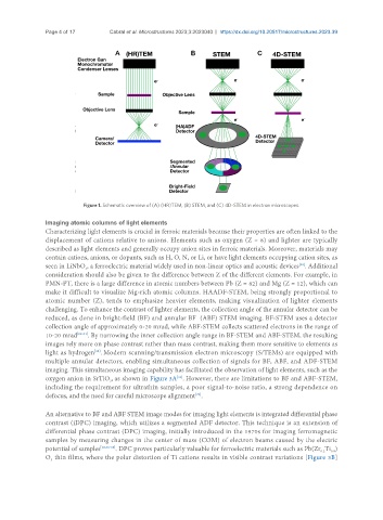
Figure 1. Schematic overview of (A) (HR)TEM, (B) STEM, and (C) 4D-STEM in electron microscopes.
Imaging atomic columns of light elements
Characterizing light elements is crucial in ferroic materials because their properties are often linked to the
displacement of cations relative to anions. Elements such as oxygen (Z = 8) and lighter are typically
described as light elements and generally occupy anion sites in ferroic materials. Moreover, materials may
contain cations, anions, or dopants, such as H, O, N, or Li, or have light elements occupying cation sites, as
[28]
seen in LiNbO , a ferroelectric material widely used in non-linear optics and acoustic devices . Additional
3
consideration should also be given to the difference between Z of the different elements. For example, in
PMN-PT, there is a large difference in atomic numbers between Pb (Z = 82) and Mg (Z = 12), which can
make it difficult to visualize Mg-rich atomic columns. HAADF-STEM, being strongly proportional to
atomic number (Z), tends to emphasize heavier elements, making visualization of lighter elements
challenging. To enhance the contrast of lighter elements, the collection angle of the annular detector can be
reduced, as done in bright-field (BF) and annular BF (ABF) STEM imaging. BF-STEM uses a detector
collection angle of approximately 0-20 mrad, while ABF-STEM collects scattered electrons in the range of
10-20 mrad [29-31] . By narrowing the inner collection angle range in BF-STEM and ABF-STEM, the resulting
images rely more on phase contrast rather than mass contrast, making them more sensitive to elements as
[32]
light as hydrogen . Modern scanning/transmission electron microscopy (S/TEMs) are equipped with
multiple annular detectors, enabling simultaneous collection of signals for BF, ABF, and ADF-STEM
imaging. This simultaneous imaging capability has facilitated the observation of light elements, such as the
oxygen anion in SrTiO , as shown in Figure 3A . However, there are limitations to BF and ABF-STEM,
[33]
3
including the requirement for ultrathin samples, a poor signal-to-noise ratio, a strong dependence on
defocus, and the need for careful microscope alignment .
[20]
An alternative to BF and ABF STEM image modes for imaging light elements is integrated differential phase
contrast (iDPC) imaging, which utilizes a segmented ADF detector. This technique is an extension of
differential phase contrast (DPC) imaging, initially introduced in the 1970s for imaging ferromagnetic
samples by measuring changes in the center of mass (COM) of electron beams caused by the electric
potential of samples [18,36-38] . DPC proves particularly valuable for ferroelectric materials such as Pb(Zr Ti )
0.2
0.8
O thin films, where the polar distortion of Ti cations results in visible contrast variations [Figure 3B]
3