
Page 172 - Read Online
P. 172
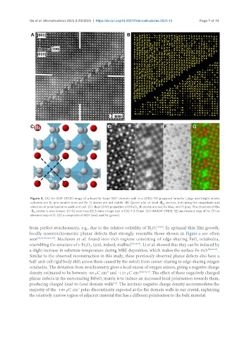
Ge et al. Microstructures 2023;3:2023026  https://dx.doi.org/10.20517/microstructures.2023.13  Page 7 of 10


                Figure 4. (A) An ADF-STEM image of a head-to-head 180° domain wall in a (010) FIB-prepared lamella. Large and bright atoms
                columns are Bi, and smaller ones are Fe. O atoms are not visible. (B) Quiver plot of local -δ  vectors, indicating the magnitude and
                                                                               FB
                direction of polarisation in each unit cell. (C) Ideal (010) projection of BiFeO ; Bi atoms are red, Fe blue, and O grey. The direction of the
                                                                  3
                -δ  vector is also shown. (D-G) core-loss EELS data: image size is 1.32 × 3.13 nm. (D) HAADF-STEM; (E) an element map of Fe; (F) an
                 FB
                element map of O; (G) a composite of ADF (red) and Fe (green).
               from perfect stoichiometry, e.g., due to the relative volatility of Bi O 3 [33,35] . In epitaxial thin film growth,
                                                                         2
               locally nonstoichiometric planar defects that strongly resemble those shown in Figure 4 are often
               seen [22,29,30,32,36,37] . Maclaren et al. found iron-rich regions consisting of edge-sharing FeO  octahedra,
                                                                                               6
               resembling the structure of γ-Fe O , (and, indeed, mullite) [30,36,37] . Li et al. showed that they can be induced by
                                          2
                                            3
               a slight increase in substrate temperature during MBE deposition, which makes the surface Fe-rich [22,29,32] .
               Similar to the observed reconstruction in this study, these previously observed planar defects also have a
               half-unit-cell rigid body shift across them caused by the switch from corner-sharing to edge-sharing oxygen
               octahedra. The deviation from stoichiometry gives a local excess of oxygen anions, giving a negative charge
               density estimated to be between -68 μC cm  and -110 μC cm -2[22,36,37] . The effect of these negatively charged
                                                    -2
               planar defects in the surrounding BiFeO  matrix is to induce an increased local polarisation towards them,
                                                  3
               producing charged head-to-head domain walls . The intrinsic negative charge density accommodates the
                                                       [32]
                                      -2
               majority of the -190 μC cm  polar discontinuity expected at the flat domain walls in our crystal, explaining
               the relatively narrow region of adjacent material that has a different polarisation to the bulk material.