
Page 65 - Read Online
P. 65
Morgan et al. Vessel Plus 2020;4:6 I http://dx.doi.org/10.20517/2574-1209.2019.32 Page 7 of 14
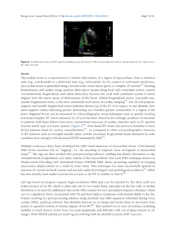
Figure 5. Transthoracic echo with RV speckle tracking based calculation of RV endocardial deformation (green vectors). RA: right atrium;
RV: right ventricle
Strain
Myocardial strain is a measurement of relative deformation of a region of myocardium, from a reference
state (e.g., end-diastole) to a deformed state (e.g., end-systole). In the context of continuum mechanics,
[42]
myocardial strain is quantified using a second-order strain tensor given its complex 3D motion . Existing
biomechanics and cardiac image analysis often report strains along local wall coordinate system, namely
circumferential, longitudinal, and radial directions, because the local wall coordinate system is better
aligned with the major mode of deformation of the heart. Global longitudinal strain, especially end-
[43]
systolic longitudinal strain, is the most commonly used metric in cardiac imaging . For clinical purpose,
negative end-systolic longitudinal strain indicates shortening of the LV with respect to end-diastole, with
more negative values indicating greater shortening and therefore greater contractility of a region of the
heart. Regional Strain can be measured by echocardiography, using techniques such as speckle tracking
and tissue Doppler; RV strain measured by 2D echo has been shown to be a stronger predictor of outcomes
in patients with heart failure than more conventional measures of cardiac function such as LV ejection
[44]
fraction and B-type natriuretic peptide [Figure 5] . Echo-based RV strain also predicts mortality in heart
[45]
failure patients listed for cardiac transplantation . As compared to other echocardiographic measures
of RV function such as tricuspid annular plane systolic excursion, longitudinal strain measured by echo
[38]
correlates most strongly with decreased RVEF measured by MRI .
Multiple techniques have been developed for MRI-based measures of myocardial strain. Conventional
MRI strain measures rely on “tagging”, i.e., the encoding of magnetic lines orthogonal to myocardial
[46]
tissue . The tags are then tracked with post-processing software, yielding fine-detail information on the
circumferential, longitudinal, and radial motion of the myocardium. One such MRI technique, known as
Displacement ENcoding with Stimulated Echoes (DENSE) MRI, shows promising capability in imaging
myocardial displacement on a voxel-by-voxel basis. This technique has been successfully applied to
[47]
examine LV motion for both human and animals under physiological and pathological conditions . Effort
[48]
has also recently been made to extend the analysis to the RV in healthy rat hearts .
All tag-based techniques require high-resolution MRI and can be limited by the thin wall and
trabeculations of the RV, which is often only one to two voxels thick, especially on the free wall. A further
limitation is the need for additional time in the MRI scanner for each specialized sequence obtained, which
can be a significant factor for patients with TR and heart failure symptoms, with limited ability to lay flat.
Feature-tracking is a post-processing solution using standard cine MRI sequences (obtained during every
cardiac MRI), applying software that identifies features in an image and tracks them in successive time
points, to quantify motion of various regions of the RV . This method has its own limitations, including
[46]
inability to track motion of less than one-pixel magnitude, and difficulty with out-of-plane motion in 2D
images. Both DENSE and feature tracking are evolving tools for detailed analysis of RV function.