
Page 61 - Read Online
P. 61
Morgan et al. Vessel Plus 2020;4:6 I http://dx.doi.org/10.20517/2574-1209.2019.32 Page 3 of 14
A B C
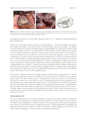
Figure 1. A: Intra-operative photograph of the tricuspid valve with closed leaflets; B: Intra-operative photograph of the tricuspid valve
with visible papillary muscles. AL: Anterior leaflet; PL: Posterior leaflet; SL: Septal leaflet; CS: Coronary sinus; TT: Tendon of Todaro; APM:
Anterior papillary muscle; PPM: Posterior papillary muscle; RA: Robot arm
physiologically function as a one-way valve. Disorders of the RV or TV disrupt the unidirectional flow of
blood, leading to TR.
The TV has three leaflets: anterior, posterior, and septal [Figure 1]. The anterior leaflet is the largest,
most mobile, and longest in the radial direction of the three leaflets. The posterior leaflet is the shortest
circumferentially and is the least consistently present; in approximately 10% of people, the posterior leaflet
[24]
and anterior leaflet are fused . The septal leaflet is the least mobile of the three leaflets and is attached
to the tricuspid annulus directly above the interventricular septum. These leaflets are suspended in a
flexible D-shaped annulus, and tethered to the RV by the sub-valvular apparatus, which is composed
of the papillary muscles and their associated chordae tendinae [Figure 1B and C]. The flexible nature of
the TV annulus allows it to adapt and change shape during the cardiac cycle, such that it can increase
[23]
up to 30% in area during end systole/early diastole . There are classically three papillary muscles: the
anterior and posterior, which are most reliably present, and the septal papillary muscle, which is absent
[25]
in approximately 20% of patients . However, the number of papillary muscles is highly variable and can
range from two to nine distinct entities. The anterior papillary muscle is the largest, and, in some patients,
the moderator band, which carries part of the right bundle branch of the atrioventricular (AV) conduction
system, can be found to join the anterior papillary muscle.
The anatomy of adjacent structures is equally important in the operative management of TV disease.
There are three important structures to consider: (1) the noncoronary sinus of Valsalva; (2) the conduction
[24]
system; and (3) the right coronary artery . The noncoronary sinus is located near the commissure
between the anterior and septal leaflets of the TV, increasing the risk of aortic perforation with
transcatheter devices, which require anchoring in this area. The AV node lies in the apex of the triangle of
Koch, and the bundle of His crosses the attachment to the septal leaflet approximately 3-5 mm posterior to
[26]
the anteroseptal commissure, which can result in heart block if there is excess pressure on the AV node .
The right coronary artery originates from the right coronary sinus of Valsalva and courses adjacent to the
tricuspid annulus, and it can result in cardiogenic shock secondary to direct injury to the right coronary
[27]
artery in TV repair .
ETIOLOGIES OF TR
TR can be divided into two overarching categories: primary and secondary. Primary TR indicates
[3]
dysfunction of the valve leaflets or chordae and makes up about 10% of TR in adults . Causes of primary
TR include: leaflet damage due to implanted devices, such as pacemakers and catheters; myxomatous
valve disease; carcinoid heart syndrome; congenital anomalies of cardiac development; endocarditis; and
rheumatic valve disease. In the absence of other cardiac disease, isolated moderate to severe primary TR
[1,3]
carries with it an estimated 1.6 × risk of mortality over the general population .