
Page 64 - Read Online
P. 64
Page 6 of 14 Morgan et al. Vessel Plus 2020;4:6 I http://dx.doi.org/10.20517/2574-1209.2019.32
A B
C
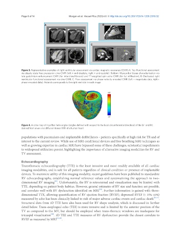
Figure 3. Representative examples of right ventricular assessment via cardiac magnetic resonance (CMR).A: Top: Functional assessment
via steady state free precession cine-CMR (left = end-diastole, right = end-systole). Bottom: Myocardial tissue characterization via
late gadolinium enhancement CMR (for infarction/fibrosis) and T1-weighted spin echo-CMR (for fat infiltration); B: Dedicated right
ventricular functional assessment via cine-CMR; C: Flow assessment via phase-velocity encoded CMR (left = magnitude data, right=
phase-encoded data). Asterisk corresponds to the right ventricle in each image
Figure 4. A color map of myofiber helix angles (angles defined with respect to the local circumferential direction) of the LV and RV,
derived from an ex vivo diffusion tensor MRI of a human heart
populations with pacemakers and implantable defibrillators - patients specifically at high risk for TR and of
interest to the current review. While use of MRI conditional devices and free breathing MRI techniques as
well as growing expertise in cardiac MRI have bypassed some of these challenges, substantial impediments
to widespread utilization persist, highlighting the importance of alternative imaging modalities for RV and
TV assessment.
Echocardiography
Transthoracic echocardiography (TTE) is the least invasive and most readily available of all cardiac
imaging modalities, and is safe for all patients regardless of clinical condition or presence of implantable
devices. To maximize utility of this imaging modality, recent guidelines have been published to standardize
RV echocardiography, establishing normal reference values and systematizing the approach to two-
[40]
dimensional RV imaging . Unfortunately, the RV is retrosternal and visualization may be limited with
TTE, depending on patient body habitus. However, general estimates of RV size and function are possible,
[35]
and correlate well with RV dysfunction identified on MRI . Further information is gained with three-
dimensional TTE, allowing quantification of RV ejection fraction (RVEF); depressed RVEF (< 35%-40%)
measured by echo has been clinically linked to risk of major adverse cardiac events and cardiac death [33,41] .
Structural data from 3D TTE have also been used for RV shape analysis, which is discussed in further
detail below. Trans-esophageal echo (TEE) is more invasive and is limited by the anterior position of the
TV as compared to the MV, but should be employed when trans-thoracic windows are inadequate for
[40]
tricuspid visualization . 3D TEE and TTE measures of RV dysfunction provide the closest correlate to
RVEF as measured by MRI [35,38] .