
Page 68 - Read Online
P. 68
Page 10 of 14 Morgan et al. Vessel Plus 2020;4:6 I http://dx.doi.org/10.20517/2574-1209.2019.32
A B
C D E
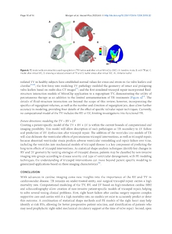
Figure 6. FE model with simulated tricuspid regurgitation (TR) before and after virtual MitraClip (MC). A: baseline mode; B: with TR jet; C:
model after virtual MC; D: showing a reduced amount of TR and E) leaflet stress after virtual MC. AL: Anterior leaflet
isolated TV in healthy subjects have established normal values for stress and strain in the valve leaflets and
chordae [18,49] ; the first foray into modeling TV pathology modeled the geometry of intact and prolapsing
[71]
valve leaflets based on multi-slice CT images ; and the first simulated tricuspid repair incorporated fluid-
structure interaction models of MitraClip application to a regurgitant TV, demonstrating the utility of
[17]
percutaneous therapy as an addition to the limited armamentarium of TR treatments [Figure 6] . The
details of fluid-structure interaction are beyond the scope of this review; however, incorporating the
specifics of regurgitant volumes, as well as the number and direction of regurgitant jets, does allow further
accuracy in modeling, providing finer details of the effect of specific valvular repair techniques. Currently,
no computational model of the TV includes the RV or LV, limiting investigations into functional TR.
Future directions: modeling the TV + RV + LV
Creating a patient-specific model of the TV + RV + LV is within the current bounds of computational and
imaging possibility. This model will allow description of such pathologies as TR secondary to LV failure
and prediction of RV dysfunction after tricuspid repair. The addition of the ventricles into models of TR
will also delineate the ventricular effects of percutaneous tricuspid interventions, as well as tricuspid repair;
because abnormal ventricular strain predicts adverse ventricular remodeling and repair failure over time,
including the ventricles into mechanical models of tricuspid disease is a key component of predicting the
long-term effects of tricuspid interventions. As statistical shape analysis techniques identify key changes in
RV and LV geometry by varying etiologies of tricuspid disease, patients may be classified by non-invasive
imaging into groups according to disease severity and type of ventricular derangement; with FE modeling
techniques, the understanding of tricuspid interventions can move beyond patient specific modeling to
[58]
generalized applications based on these imaging characteristics .
CONCLUSION
With advances in cardiac imaging come new insights into the importance of the RV and TV in
cardiovascular disease. TR remains an under-treated entity, and surgical tricuspid repair carries a high
mortality rate. Computational modeling of the TV, RV, and LV based on high-resolution cardiac MRI
and echocardiography allow creation of non-invasive patient-specific models of tricuspid repair, helping
to solve several vexing clinical problems. First, right heart failure after cardiac surgery requires complex
supportive care and carries with it a high mortality rate; no models yet exist to accurately predict or deter
this outcome. A combination of statistical shape methods and FE models of the right heart may help
identify at-risk RVs, allowing for better preoperative patient selection, and identification of patients who
may need prophylactic right-sided mechanical circulatory support at the time of valve repair. Second, open