
Page 95 - Read Online
P. 95
Shalimova Cardiovascular remodeling in EH and DM2
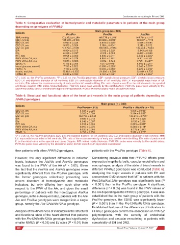
Table 5: Comparative evaluation of hemodynamic and metabolic parameters in patients of the main group
depending on genotypes of PPARγ2
Main group (n = 320)
Indices
Pro/Pro Pro/Ala Ala/Ala
SBP, mmHg 172.372 ± 0.259 165.775 ± 0.351* 165.714 ± 1.017*
DBP, mmHg 101.599 ± 0.196 99.338 ± 0.232* 100.001 ± 1113
EDD LV, cm 5.024 ± 0.026 4.878 ± 0.038* 4.856 ± 0.084
ESD LV, cm 3.316 ± 0.024 3.198 ± 0.034* 3.149 ± 0.072
MMI LV, g/m 142.794 ± 2.551 136.553 ± 2.983 139.933 ± 7.664
E/A 0.924 ± 0.013 0.915 ± 0.027 0.943 ± 0.158
E/e 6.348 ± 0.097 6.108 ± 0.193 6.422 ± 0.866
IMT, mm 0.951 ± 0.006 0.900 ± 0.011* 0.849 ± 0.035*
PWV of the CA, m/c 8.910 ± 0.075 8.592 ± 0.122* 8.301 ± 0.301
PWV of the AA, m/c 9.020 ± 0.086 8.874 ± 0.146 7.779 ± 0.461*º
EDVD, % 6.155 ± 0.056 7.017 ± 0.079* 6.959 ± 0.241*
blood glucose, mmol/L 7.190 ± 0.022 6.968 ± 0.029* 6.643 ± 0.057*
HbA1c, % 7.103 ± 0.013 6.939 ± 0.058* 6.929 ± 0.042*
insulin, mcU/mL 25.182 ± 0.255 21.906 ± 1.526 22.814 ± 1.735
HOMA-IR 8.039 ± 0.083 6.767 ± 0.156* 6.722 ± 0.484*
*P < 0.05 vs. the Pro/Pro genotypes; ºP < 0.05 vs. the Pro/Ala genotypes. SBP: systolic blood pressure; DBP: diastolic blood pressure;
EDD LV: end-diastolic diameter of left ventricle; ESD LV: end-systolic diameter of left ventricle; MMI LV: myocardial mass index of left
ventricle; E/A: ratio of the maximum velocity of early and late left ventricle filling; E/e: ratio of peak e and E on the mitral valve in the spectral
and tissue Doppler; IMT: intima media thickness; PVW CA: pulse wave velocity by the carotid artery; PVW AA: pulse wave velocity by the
abdominal aortic; EDVD: endothelium dependent vasodilation; HOMA-IR: homeostasis model assessment index
Table 6: Structural and functional state of the heart and vessels in the main group of patients depending on
PPARγ2 genotypes
Main group (n = 320)
Indices
Pro/Pro (n = 242) Pro/Ala + Ala/Ala (n = 78)
EDD LV, cm 5.024 ± 0.026 4.876 ± 0.035*
ESD LV, cm 3.316 ± 0.024 3.194 ± 0.032*
MMI LV, g/m 142.794 ± 2.551 133.215 ± 2.799*
E/A 0.924 ± 0.013 0.917 ± 0.028
E/e 6.348 ± 0.097 6.136 ± 0.190
IMT, mm 0.951 ± 0.006 0.895 ± 0.011*
PWV of the CA, m/c 8.910 ± 0.075 8.566 ± 0.114*
PWV of the AA, m/c 9.020 ± 0.086 8.776 ± 0.143
EDVD, % 6.155 ± 0.056 7.012 ± 0.075*
*P < 0.05 vs. the Pro/Pro genotypes. EDD LV: end-diastolic diameter of left ventricle; ESD LV: end-systolic diameter of left ventricle; MMI
LV: myocardial mass index of left ventricle; E/A: ratio of the maximum velocity of early and late left ventricle filling; E/e: ratio of peak e and
E on the mitral valve in the spectral and tissue Doppler; IMT: intima media thickness; PVW CA: pulse wave velocity by the carotid artery;
PVW AA: pulse wave velocity by the abdominal aortic; EDVD: endothelium dependent vasodilation
than patients with other PPARγ2 genotypes. patients with the Pro/Pro genotype [Table 6].
However, the only significant difference in indicator Considering previous data that PPARγ2 affects gene
levels, between the Ala/Ala and Pro/Ala genotypes expression in epithelial cells, vascular endothelium and
was found in the PWV of the AA (P < 0.05). Given macrophages, analysis of the state of blood vessels in
the fact that the Pro/Ala and Ala/Ala genotypes were different PPARγ2 genotypes was conducted [Table 6].
significantly different from the Pro/Pro genotype, with Analyzing the major vessels in patients with EH and
the former genotypes collectively presenting less concomitant DM2 showed that IMT in patients with the
severe disorders of hemodynamic and metabolic Pro12Ala/Ala12Ala genotype was significantly less (P
indicators, but only differing from each other with < 0.001) than in the Pro/Pro genotype. A significant
respect to the PWV of the AA, and given the small difference (P < 0.05) was found in the PWV values of
percentage of patients with the homozygous Ala/Ala the CA depending on the PPARγ2 genotype. It was also
genotype, in the subsequent step, patients with the Ala/ established that in the main group of patients with the
Ala and Pro/Ala genotypes were merged into a single Pro/Pro genotype, the EDVD was significantly lower
group, namely the Pro12Ala/Ala12Ala genotype. (P < 0.001) than in the Pro12Ala/Ala12Ala genotype.
Established features of the differences of indicators in
Analysis of the differences of indicators in the structural PPARγ2 genotypes confirm the association of PPARγ2
and functional state of the heart showed that patients polymorphisms with the severity of endothelial
with the Pro12Ala/Ala12Ala genotype had significantly dysfunction and vascular remodeling in patients with
smaller MMILV (P < 0.05) and LV sizes (P < 0.01) than comorbidity of EH and DM2.
88 Vessel Plus ¦ Volume 1 ¦ June 27, 2017