
Page 18 - Read Online
P. 18
Rao. Vessel Plus 2022;6:22 https://dx.doi.org/10.20517/2574-1209.2021.105 Page 7 of 24
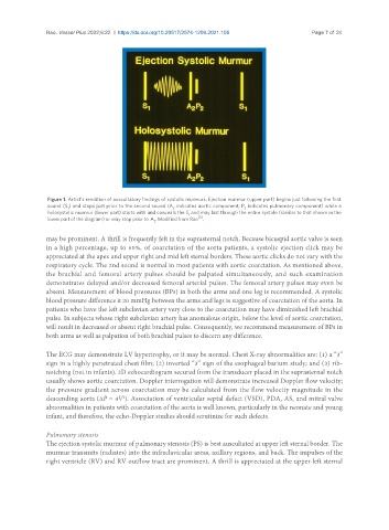
Figure 1. Artist’s rendition of auscultatory findings of systolic murmurs. Ejection murmur (upper part) begins just following the first
sound (S ) and stops just prior to the second sound (A indicates aortic component; P indicates pulmonary component) while a
1 2 2
holosystolic murmur (lower part) starts with and conceals the S and may last through the entire systole (similar to that shown in the
1
[5]
lower part of the diagram) or may stop prior to A . Modified from Rao .
2
may be prominent. A thrill is frequently felt in the suprasternal notch. Because bicuspid aortic valve is seen
in a high percentage, up to 60%, of coarctation of the aorta patients, a systolic ejection click may be
appreciated at the apex and upper right and mid left sternal borders. These aortic clicks do not vary with the
respiratory cycle. The 2nd sound is normal in most patients with aortic coarctation. As mentioned above,
the brachial and femoral artery pulses should be palpated simultaneously, and such examination
demonstrates delayed and/or decreased femoral arterial pulses. The femoral artery pulses may even be
absent. Measurement of blood pressures (BPs) in both the arms and one leg is recommended. A systolic
blood pressure difference ≥ 20 mmHg between the arms and legs is suggestive of coarctation of the aorta. In
patients who have the left subclavian artery very close to the coarctation may have diminished left brachial
pulse. In subjects whose right subclavian artery has anomalous origin, below the level of aortic coarctation,
will result in decreased or absent right brachial pulse. Consequently, we recommend measurement of BPs in
both arms as well as palpation of both brachial pulses to discern any difference.
The ECG may demonstrate LV hypertrophy, or it may be normal. Chest X-ray abnormalities are: (1) a “3”
sign in a highly penetrated chest film; (2) inverted “3” sign of the esophageal barium study; and (3) rib-
notching (not in infants). 2D echocardiogram secured from the transducer placed in the suprasternal notch
usually shows aortic coarctation. Doppler interrogation will demonstrate increased Doppler flow velocity;
the pressure gradient across coarctation may be calculated from the flow velocity magnitude in the
2
descending aorta (ΔP = 4V ). Association of ventricular septal defect (VSD), PDA, AS, and mitral valve
abnormalities in patients with coarctation of the aorta is well known, particularly in the neonate and young
infant, and therefore, the echo-Doppler studies should scrutinize for such defects.
Pulmonary stenosis
The ejection systolic murmur of pulmonary stenosis (PS) is best auscultated at upper left sternal border. The
murmur transmits (radiates) into the infraclavicular areas, axillary regions, and back. The impulses of the
right ventricle (RV) and RV outflow tract are prominent. A thrill is appreciated at the upper left sternal