
Page 14 - Read Online
P. 14
Page 4 of 24 Rao. Vessel Plus 2022;6:22 https://dx.doi.org/10.20517/2574-1209.2021.105
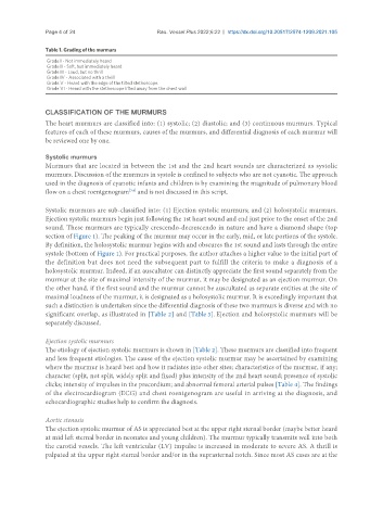
Table 1. Grading of the murmurs
Grade I - Not immediately heard
Grade II - Soft, but immediately heard
Grade III - Loud, but no thrill
Grade IV - Associated with a thrill
Grade V - Heard with the edge of the tilted stethoscope
Grade VI - Heard with the stethoscope lifted away from the chest wall
CLASSIFICATION OF THE MURMURS
The heart murmurs are classified into: (1) systolic; (2) diastolic; and (3) continuous murmurs. Typical
features of each of these murmurs, causes of the murmurs, and differential diagnosis of each murmur will
be reviewed one by one.
Systolic murmurs
Murmurs that are located in between the 1st and the 2nd heart sounds are characterized as systolic
murmurs. Discussion of the murmurs in systole is confined to subjects who are not cyanotic. The approach
used in the diagnosis of cyanotic infants and children is by examining the magnitude of pulmonary blood
[3,4]
flow on a chest roentgenogram and is not discussed in this script.
Systolic murmurs are sub-classified into: (1) Ejection systolic murmurs; and (2) holosystolic murmurs.
Ejection systolic murmurs begin just following the 1st heart sound and end just prior to the onset of the 2nd
sound. These murmurs are typically crescendo-decrescendo in nature and have a diamond shape (top
section of Figure 1). The peaking of the murmur may occur in the early, mid, or late portions of the systole.
By definition, the holosystolic murmur begins with and obscures the 1st sound and lasts through the entire
systole (bottom of Figure 1). For practical purposes, the author attaches a higher value to the initial part of
the definition but does not need the subsequent part to fulfill the criteria to make a diagnosis of a
holosystolic murmur. Indeed, if an auscultator can distinctly appreciate the first sound separately from the
murmur at the site of maximal intensity of the murmur, it may be designated as an ejection murmur. On
the other hand, if the first sound and the murmur cannot be auscultated as separate entities at the site of
maximal loudness of the murmur, it is designated as a holosystolic murmur. It is exceedingly important that
such a distinction is undertaken since the differential diagnosis of these two murmurs is diverse and with no
significant overlap, as illustrated in [Table 2] and [Table 3]. Ejection and holosystolic murmurs will be
separately discussed.
Ejection systolic murmurs
The etiology of ejection systolic murmurs is shown in [Table 2]. These murmurs are classified into frequent
and less frequent etiologies. The cause of the ejection systolic murmur may be ascertained by examining
where the murmur is heard best and how it radiates into other sites; characteristics of the murmur, if any;
character (split, not split, widely split and fixed) plus intensity of the 2nd heart sound; presence of systolic
clicks; intensity of impulses in the precordium; and abnormal femoral arterial pulses [Table 4]. The findings
of the electrocardiogram (ECG) and chest roentgenogram are useful in arriving at the diagnosis, and
echocardiographic studies help to confirm the diagnosis.
Aortic stenosis
The ejection systolic murmur of AS is appreciated best at the upper right sternal border (maybe better heard
at mid left sternal border in neonates and young children). The murmur typically transmits well into both
the carotid vessels. The left ventricular (LV) impulse is increased in moderate to severe AS. A thrill is
palpated at the upper right sternal border and/or in the suprasternal notch. Since most AS cases are at the