Page 16 - Read Online
P. 16

Page 6 of 24                                                                                            Rao. Vessel Plus 2022;6:22    https://dx.doi.org/10.20517/2574-1209.2021.105


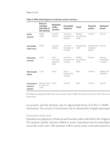
                              Table 4. Differential diagnosis of ejection systolic murmurs

                                           Point of      Radiation                                                        Ejection
                                           maximal       of the       Precordial   Thrill        Femoral     2nd heart    systolic     Chest X-ray     ECG            Echo-Doppler         Other features
                                           intensity of   murmur      impulses                   pulses      sound        click
                                           the murmur
                              Aortic       RUSB          Carotid      Normal or    RUSB &        Normal      Normal       Constant     Dilated ascending  Normal or LVH  Thickened bicuspid   Severity of aortic stenosis
                              stenosis     LMSB          arteries     increased LV  suprasternal                          click at apex,  aorta                       aortic valve leaflets,   is difficult to judge by
                                                                      impulse      notch                                  LMSB &                                      increased Doppler flow  clinical examination
                                                                                                                          RUSB                                        velocity across the
                                                                                                                                                                      aortic valve
                              Coarctation   RUSB         Carotid      Normal or    Suprasternal   Decreased &  Normal     Constant     Inverted 3 sign on  Normal or left   Suprasternal notch 2D  Measurement of blood
                              of the aorta               arteries     increased LV  notch        delayed or               click at apex,  barium-filled   ventricular   echo shows         pressure in arms and legs
                                                                      impulse                    absent                   LMSB &       esophagus, rib   hypertrophy   coarctation, increased  is helpful
                                                                                                                          RUSB         notching        (LVH)          flow velocity in
                                                                                                                                                                      descending aorta
                              Pulmonary    LUSB          Infra-       Normal or    LSUB &        Normal      Normal,      LUSB         Dilated main    Normal or RVH RV enlargement,       Duration & timing of
                              stenosis                   clavicular   increased RV  suprasternal             diminished,   LMSB        pulmonary artery               increased Doppler flow  peaking of the murmur,
                                                         regions &    impulse      notch                     or absent    LLSB, varies                                velocity across the   degree of splitting &
                                                         back                                                             with                                        pulmonary valve      intensity of 2nd sound may
                                                                                                                          respiration                                                      suggest severity of
                                                                                                                                                                                           stenosis
                              Atrial septal   LUSB       None         Hyper-       None          Normal      Widely split   None       Prominent main   Mild RVH      Enlarged RV,         Mid-diastolic murmur at
                              defect                                  dynamic RV                             and fixed                 pulmonary artery,              paradoxical septal   LLSB
                                                                      impulse                                                          increased                      motion, atrial defect on
                                                                                                                                       pulmonary blood                subcostal echo-
                                                                                                                                       flow                           Doppler

                              Functional or   Between apex  None      Normal       None          Normal      Normal       None         Normal          Normal         Normal               Vibratory or musical
                              innocent     & LLSB or at                                                                                                                                    quality to the murmur
                              murmur       LUSB


                              ECG: Electrocardiogram; RUSB: right upper sternal border; LMSB: left mid-sternal border; LLSB: left lower sternal border; LUSB: left upper sternal border; LV: left ventricular; RV: right ventricle; RVH: right ventricular
                              hypertrophy.


                              are present, and the murmur may be appreciated better at LLSB or LMSBs. However, 2D echo studies are useful in defining the site of LV outflow tract
                              obstruction. The severity of obstruction can be estimated by Doppler interrogation of the LV outflow tract and supravalvar aortic region.


                              Coarctation of the aorta
                              Simultaneous palpation of femoral and brachial pulses will lead to the diagnosis of coarctation of the aorta, although the murmur is the presenting complaint.
                              The ejection systolic murmur related to aortic coarctation may be auscultated at the right upper sternal border and is probably caused by flow disturbance

                              across the aortic valve. The murmur of flow across aortic coarctation may be auscultated best in the left inter-scapular region over the back. The LV impulse
   11   12   13   14   15   16   17   18   19   20   21