
Page 20 - Read Online
P. 20
Rao. Vessel Plus 2022;6:22 https://dx.doi.org/10.20517/2574-1209.2021.105 Page 9 of 24
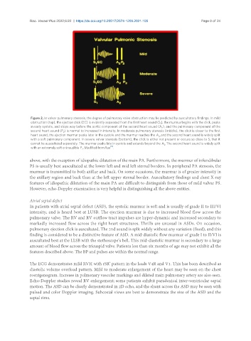
Figure 2. In valvar pulmonary stenosis, the degree of pulmonary valve obstruction may be predicted by auscultatory findings. In mild
obstruction (top), the ejection click (EC) is evidently separated from the first heart sound (S ), the murmur begins with the click, peaks
1
in early systole, and stops way before the aortic component of the second heart sound (A ), and the pulmonary component of the
2
second heart sound (P ) is normal to increased in intensity. In moderate pulmonary stenosis (middle), the click is closer to the first
2
heart sound, the ejection murmur peaks later in the systole and the murmur reaches the A , and the second heart sound is widely split
2
with a soft pulmonary component. In severe valvar stenosis (bottom), the click is either not present or occurs so close to S that it
1
cannot be auscultated separately. The murmur peaks late in systole and extends beyond the A . The second heart sound is widely split
2
[5]
with an extremely soft or inaudible P . Modified from Rao .
2
above, with the exception of idiopathic dilatation of the main PA. Furthermore, the murmur of infundibular
PS is usually best auscultated at the lower left and mid left sternal borders. In peripheral PA stenosis, the
murmur is transmitted to both axillae and back. On some occasions, the murmur is of greater intensity in
the axillary region and back than at the left upper sternal border. Auscultatory findings and chest X-ray
features of idiopathic dilatation of the main PA are difficult to distinguish from those of mild valvar PS.
However, echo-Doppler examination is very helpful in distinguishing all the above entities.
Atrial septal defect
In patients with atrial septal defect (ASD), the systolic murmur is soft and is usually of grade II to III/VI
intensity, and is heard best at LUSB. The ejection murmur is due to increased blood flow across the
pulmonary valve. The RV and RV outflow tract impulses are hyper-dynamic and increased secondary to
markedly increased flow across the right heart structures. Thrills are unusual in ASDs. On occasion,
pulmonary ejection click is auscultated. The 2nd sound is split widely without any variation (fixed), and this
finding is considered to be a distinctive feature of ASD. A mid-diastolic flow murmur of grade I to II/VI is
auscultated best at the LLSB with the stethoscope’s bell. This mid-diastolic murmur is secondary to a large
amount of blood flow across the tricuspid valve. Patients less than six months of age may not exhibit all the
features described above. The BP and pulses are within the normal range.
The ECG demonstrates mild RVH with rSR’ pattern in the leads V4R and V1. This has been described as
diastolic volume overload pattern. Mild to moderate enlargement of the heart may be seen on the chest
roentgenogram. Increase in pulmonary vascular markings and dilated main pulmonary artery are also seen.
Echo-Doppler studies reveal RV enlargement; some patients exhibit paradoxical inter-ventricular septal
motion. The ASD can be clearly demonstrated in 2D echo, and the shunt across the ASD may be seen with
pulsed and color Doppler imaging. Subcostal views are best to demonstrate the size of the ASD and the
septal rims.