


Page 14 - Read Online
P. 14
Lust et al. Vessel Plus 2021;5:7  I  http://dx.doi.org/10.20517/2574-1209.2020.52                                                     Page 9 of 14


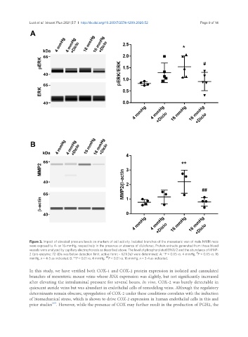
               Figure 3. Impact of elevated pressure levels on markers of cell activity. Isolated branches of the mesenteric vein of male NMRI mice
               were exposed to 4- or 16-mmHg, respectively in the presence or absence of diclofenac. Protein extracts generated from these blood
               vessels were analyzed by capillary electrophoresis as described above. The level of phosphorylated ERK1/2 and the abundance of MMP-
                                                                                               #
               2 (pro-enzyme: 72 kDa was below detection limit, active form: ~ 62 kDa) were determined; A: *P < 0.05 vs. 4 mmHg,  P < 0.05 vs. 16
                                                    ##
               mmHg, n = 4-5 as indicated; B: **P < 0.01 vs. 4 mmHg,  P < 0.01 vs. 16 mmHg, n = 3-4 as indicated.
               In this study, we have verified both COX-1 and COX-2 protein expression in isolated and cannulated
               branches of mesenteric mouse veins whose RNA expression was slightly, but not significantly increased
               after elevating the intraluminal pressure for several hours. In vivo, COX-2 was barely detectable in
               quiescent auricle veins but was abundant in endothelial cells of remodeling veins. Although the regulatory
               determinants remain obscure, upregulation of COX-2 under these conditions correlates with the induction
               of biomechanical stress, which is shown to drive COX-2 expression in human endothelial cells in this and
               prior studies . However, while the presence of COX may further result in the production of PGH2, the
                          [15]

