


Page 9 - Read Online
P. 9
Page 4 of 14                                                      Lust et al. Vessel Plus 2021;5:7  I  http://dx.doi.org/10.20517/2574-1209.2020.52

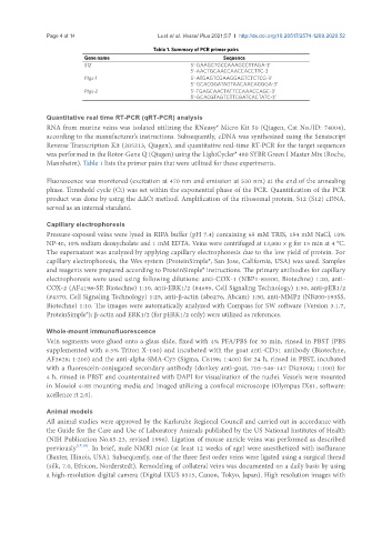
                                                Table 1. Summary of PCR primer pairs
                           Gene name                                 Sequence
                           S12                             5’-GAAGCTGCCAAAGCCTTAGA-3’
                                                           5’-AACTGCAACCAACCACCTTC-3
                           Ptgs-1                          5’-ATGAGTCGAAGGAGTCTCTCG-3’
                                                           5’-GCACGGATAGTAACAACAGGGA-3’
                           Ptgs-2                          5’-TGAGCAACTATTCCAAACCAGC-3’
                                                           5’-GCACGTAGTCTTCGATCACTATC-3’

               Quantitative real time RT-PCR (qRT-PCR) analysis
               RNA from murine veins was isolated utilizing the RNeasy® Micro Kit 50 (Qiagen, Cat No./ID: 74004),
               according to the manufacturer’s instructions. Subsequently, cDNA was synthesized using the Sensiscript
               Reverse Transcription Kit (205213, Qiagen), and quantitative real-time RT-PCR for the target sequences
               was performed in the Rotor-Gene Q (Qiagen) using the LightCycler® 480 SYBR Green I Master Mix (Roche,
               Mannheim). Table 1 lists the primer pairs that were utilized for these experiments.

               Fluorescence was monitored (excitation at 470 nm and emission at 530 nm) at the end of the annealing
               phase. Threshold cycle (Ct) was set within the exponential phase of the PCR. Quantification of the PCR
               product was done by using the ΔΔCt method. Amplification of the ribosomal protein, S12 (S12) cDNA,
               served as an internal standard.


               Capillary electrophoresis
               Pressure-exposed veins were lysed in RIPA buffer (pH 7.4) containing 65 mM TRIS, 154 mM NaCl, 10%
               NP-40, 10% sodium deoxycholate and 1 mM EDTA. Veins were centrifuged at 13,000 × g for 15 min at 4 °C.
               The supernatant was analyzed by applying capillary electrophoresis due to the low yield of protein. For
               capillary electrophoresis, the Wes system (ProteinSimple®, San Jose, California, USA) was used. Samples
               and reagents were prepared according to ProteinSimple® instructions. The primary antibodies for capillary
               electrophoresis were used using following dilutions: anti-COX-1 (NBP1-85500, Biotechne) 1:20, anti-
               COX-2 (AF4198-SP, Biotechne) 1:10, anti-ERK1/2 (#4695, Cell Signaling Technology) 1:50, anti-pER1/2
               (#4370, Cell Signaling Technology) 1:25, anti-β-actin (ab6276, Abcam) 1:50, anti-MMP2 (NB200-193SS,
               Biotechne) 1:10. The images were automatically analyzed with Compass for SW software (Version 3.1.7,
               ProteinSimple®); β-actin and ERK1/2 (for pERK1/2 only) were utilized as references.


               Whole-mount immunofluorescence
               Vein segments were glued onto a glass slide, fixed with 4% PFA/PBS for 30 min, rinsed in PBST (PBS
               supplemented with 0.5% Triton X-100) and incubated with the goat anti-CD31 antibody (Biotechne,
               AF3628; 1:200) and the anti-alpha-SMA-Cy3 (Sigma, C6198; 1:400) for 24 h, rinsed in PBST, incubated
               with a fluorescein-conjugated secondary antibody (donkey anti-goat, 705-546-147 Dianova; 1:100) for
               4 h, rinsed in PBST and counterstained with DAPI for visualization of the nuclei. Vessels were mounted
               in Mowiol 4-88 mounting media and imaged utilizing a confocal microscope (Olympus IX81, software:
               xcellence rt 2.0).

               Animal models
               All animal studies were approved by the Karlsruhe Regional Council and carried out in accordance with
               the Guide for the Care and Use of Laboratory Animals published by the US National Institutes of Health
               (NIH Publication No.85-23, revised 1996). Ligation of mouse auricle veins was performed as described
               previously [17,18] . In brief, male NMRI mice (at least 12 weeks of age) were anesthetized with isoflurane
               (Baxter, Illinois, USA). Subsequently, one of the three first order veins were ligated using a surgical thread
               (silk, 7.0, Ethicon, Norderstedt). Remodeling of collateral veins was documented on a daily basis by using
               a high-resolution digital camera (Digital IXUS 8515, Canon, Tokyo, Japan). High resolution images with

